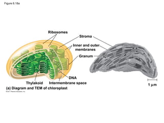
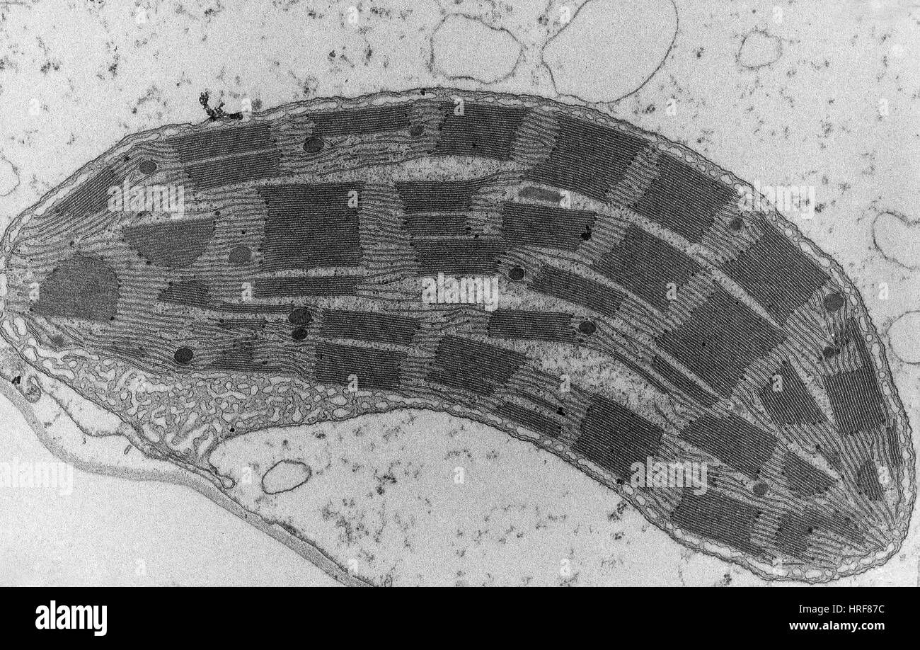
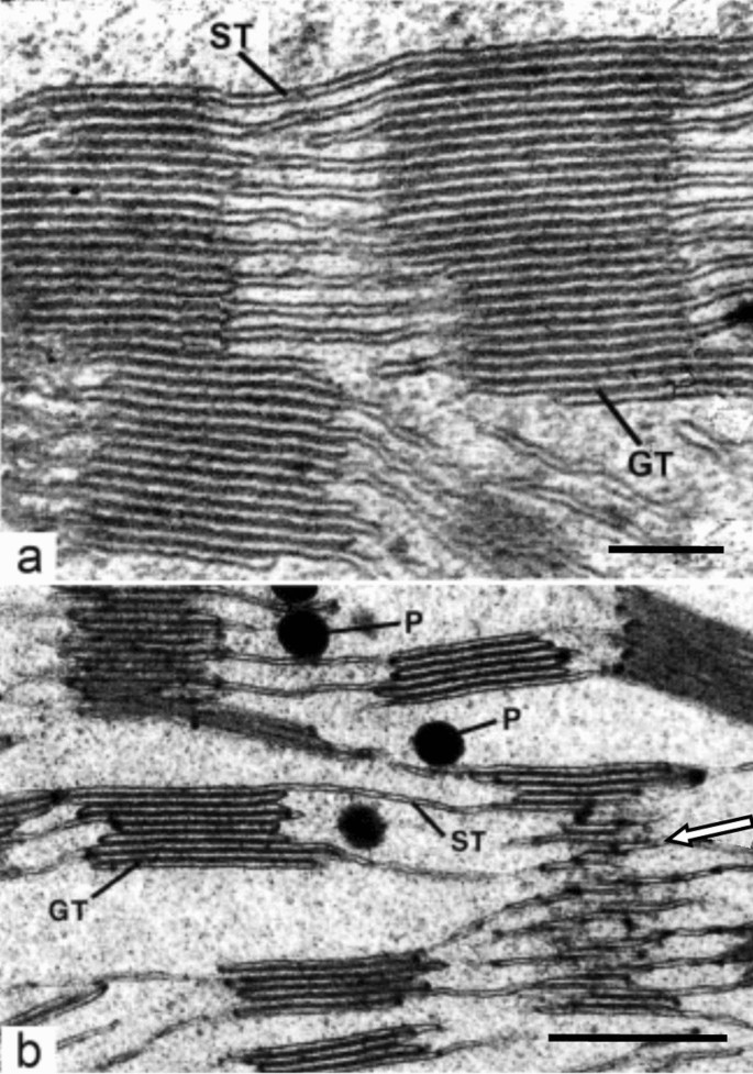
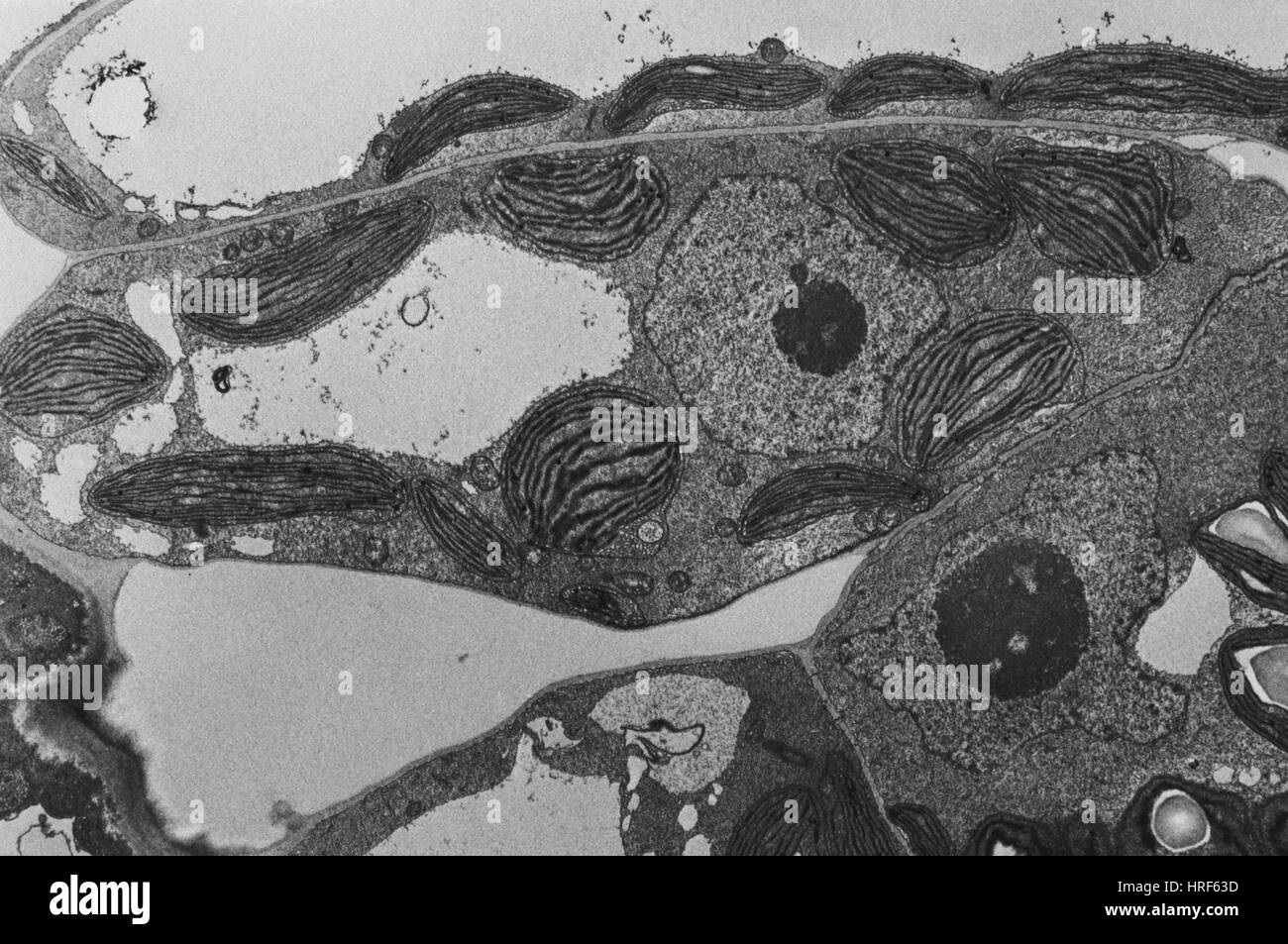
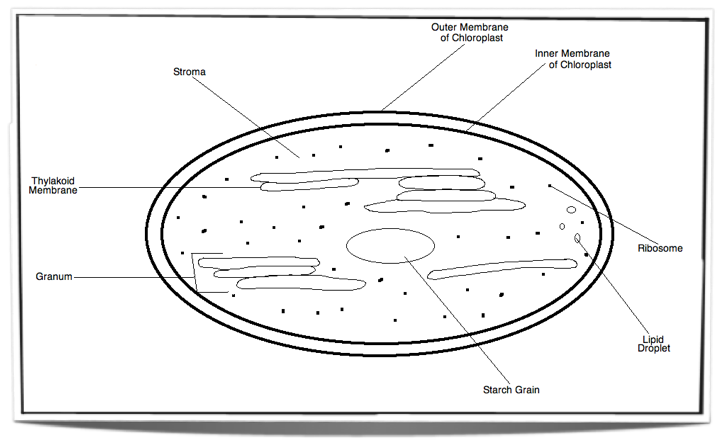
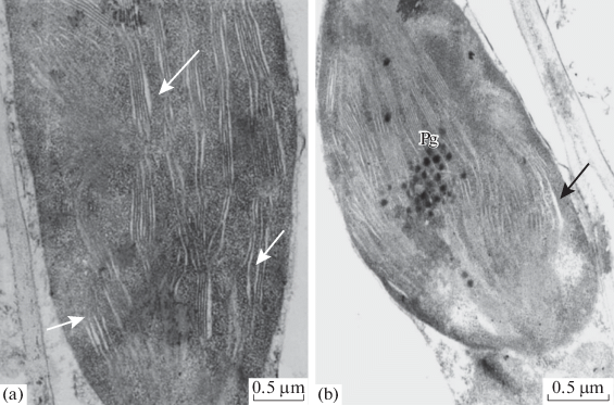
41 tem image of chloroplast labeled
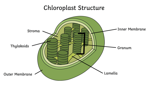
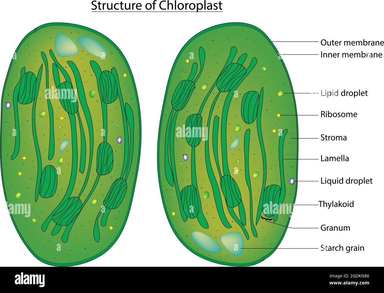
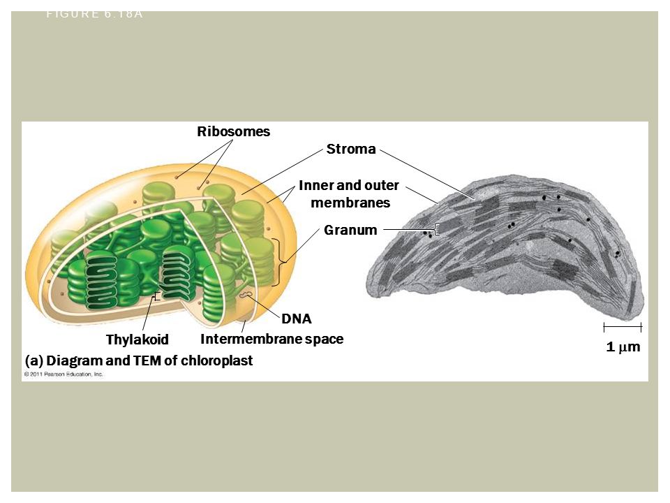
PDF Ch.06A Tour of the Cell - Estrella Mountain Community College Chloroplast Peroxisome Two subunits made of ribo- somal RNA and proteins; can be free in cytosol or bound to ER Extensive network of membrane-bound tubules and sacs; membrane separates lumen from cytosol; continuous with the nuclear envelope. Membranous sac of hydrolytic enzymes (in animal cells) Large membrane-bounded vesicle in plants Chloroplast- Diagram, Structure and Function Of Chloroplast - BYJUS Diagram of Chloroplast The chloroplast diagram below represents the chloroplast structure mentioning the different parts of the chloroplast. The parts of a chloroplast such as the inner membrane, outer membrane, intermembrane space, thylakoid membrane, stroma and lamella can be clearly marked out.
Label This Transmission Electron Micrograph : TEM of chloroplast from ... Transmission electron microscopy (tem) is a microscopy technique in which a beam of electrons is transmitted through a specimen to form an image. Label the transmission electron micrograph of the nucleus. Subset of labeled images and transfer labels to the entire image corpus. Labeling for electron microscopy using antibody conjugated to.
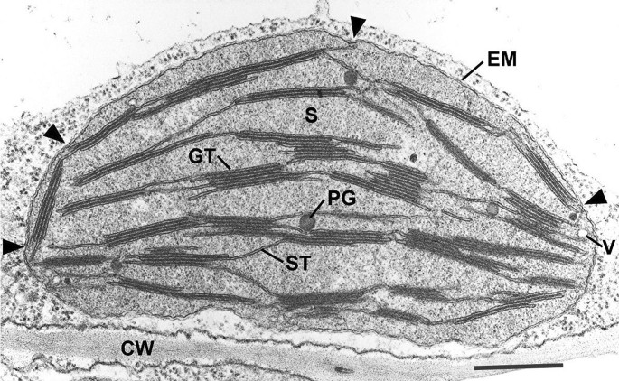
Tem image of chloroplast labeled
Electron Microscopy Images - Dartmouth Sep 10, 2021 ... Transmission electron microscope image of a thin section cut through the bronchiolar epithelium of the lung(mouse), which consists of ... Structure of Chloroplast (With Diagram) | Botany - Biology Discussion This will help you to draw the structure and diagram of chloroplast. 1. Chloroplasts (Figs. 295-296), responsible for the photosynthesis of the plants, are the characteristic features of the cells of green plants. ADVERTISEMENTS: 2. Around the chloroplast is present a double membrane envelope. 3. Each membrane of chloroplasst is 35 to 50 Å thick. PDF Plant Anatomy: Images and diagrams to explain concepts 1.6 CHLOROPLASTS Chloroplasts are large organelles and their function is the formation and storage of carbohydrates from photosynthesis. The chloroplast is bounded by a double membrane. The matrix of the chloroplast is known as the stroma. Also inside the chloroplast are separate internal membranes that form lamellae or rounded tongue-like
Tem image of chloroplast labeled. Assignment 6, page 1 - North Carolina State University Now, it is time to view a chloroplast by transmission electron microscopy. View this transmission electron micrograph of a plant cell, locate a chloroplast and capture the image for labeling.The micrograph is displayed as if using a "virtual electron microscope", so you will need to magnify the image and move to a region that contains the clearest view of chloroplast internal structures. TEM of Dynamic Autophagic Activity in Leaves during the Night. (A ... (A) Representative TEM images of autophagic structures at different time points after dark treatment. Lots of autophagic bodies (blue arrows) appeared in the central vacuole of mesophyll cells... Chloroplast Function in Photosynthesis - ThoughtCo Chlorophyll is a green photosynthetic pigment within the chloroplast grana that absorbs light energy for photosynthesis. Chloroplasts are found in plant leaves surrounded by guard cells. These cells open and close tiny pores allowing for the gas exchange needed for photosynthesis. Measuring the dynamic response of the thylakoid architecture in ... Nov 5, 2020 ... Schematic diagram of the methodology to analyze the TEM images. 1. An intact chloroplast from HPF‐treated leaf tissue is selected; 2.
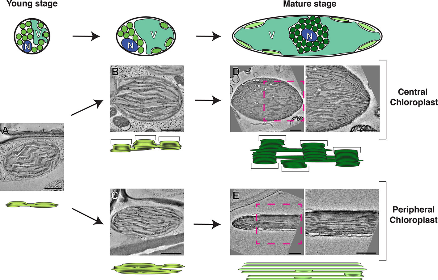
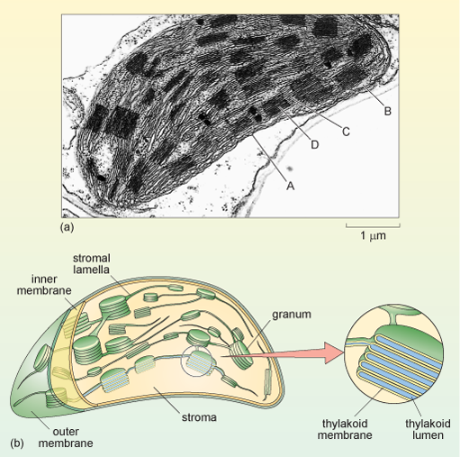
Three-Dimensional Analysis of Chloroplast Structures Associated with ... Chloroplasts are responsible for the eukaryotic photosynthesis and carbon fixation, thus providing energy for much of life on the earth. Chloroplast biogenesis is a complex process and is highly integrated with cellular and plant development (Yang et al., 2010; Pogson et al., 2015).Although three-dimensional (3D) models of the thylakoid membrane architecture have been created using electron ... Chloroplasts - Definition, Structure, Function and Microscopy To view chloroplasts under the microscope, students can use toluidine blue stain to prepare a wet mount. This simply involves the following simple steps: Place a plant sample onto drop of water on a clean glass slide Using a dropper, add a drop of the stain (toluidine blue) on the sample and allow to stand for about a minute Solved Examine this electron micrograph of a chlorplast. - Chegg Biology questions and answers. Examine this electron micrograph of a chlorplast. A. Identify the stack of membranes labeled A. B. Identify the region labeled B. C. Would the production of organic compounds during the light-independent reactions occur in. Question: Examine this electron micrograph of a chlorplast. A. Chloroplast: Meaning, Structure, Analogy - Embibe The word chloroplast comes from the Greek words 'khloros', meaning "green", and 'plastes', meaning "formed". The diagram of Chloroplast is given below. Fig: A Labeled Diagram of Chloroplast Chloroplast Structure Chloroplasts are roughly \ (1 - 2\, {\rm {μm}}\) thick and \ (5 - 7\, {\rm {μm}}\) in diameter and are seen in all higher plants.
Lab #8 photosynthesis with some questions from mito,meio an cyto - Quizlet Which will show if the leaf is conducting photosynthesis. 5) Q: would you illuminate your house plants with a green light bulb why or why not. A: No because a green lights color is not absorbed by plants it is reflected. 6) Examine this electron micrograph of a chloroplast. a) Identify the stack of membranes labeled A. Transmission electron microscopic images of chloroplasts and ... For easy organelle identification, a chloroplast (P) and a mitochondrion (M) are labeled. (C-E) Ultrastructure of chloroplasts and mitochondria in cells of the strong PRORP1 RNAi mutant line... Chloroplast Stock Photos, Pictures & Royalty-Free Images - iStock Browse 36,707 chloroplast stock photos and images available, or search for chloroplast structure or chloroplast micrograph to find more great stock photos and pictures. Chloroplast, plant cell organelle. Chloroplast, plant cell organelle. 3d image isolated on white. Round, green chloroplasts in plant cells of anacharis or waterweed, Egeria densa. Electron Microscopy Views of Dimorphic Chloroplasts in C4 Plants Jun 22, 2020 ... Advances in electron microscopy imaging capacity and sample preparation ... It is difficult to capture dividing chloroplasts by TEM because ...
Chloroplast - Wikipedia Chloroplasts visible in the cells of Bryum capillare, a type of moss A chloroplast / ˈklɔːrəˌplæst, - plɑːst / [1] [2] is a type of membrane-bound organelle known as a plastid that conducts photosynthesis mostly in plant and algal cells.
Chloroplast hi-res stock photography and images - Alamy Find the perfect chloroplast stock photo, image, vector, illustration or 360 image. ... Diagram showing chloroplast in plant leaf illustration Stock Vector.
A multifaceted analysis reveals two distinct phases of chloroplast ... Mar 3, 2021 ... Transmission electron microscopy (TEM) images of cotyledon cells of 3- day-old, dark-grown Arabidopsis thaliana (Columbia) seedlings illuminated ...
Chloroplasts | Photoreceptor Apparata | Algae - Biocyclopedia Transmission electron microscopy image of a chloroplast at higher magnification showing the thylakoid membrane and the eyespot globules (b). (Bar: 1 µm.) FIGURE 2.79 Transmission electron microscopy image of Nannochloropsis sp. in transverse section, showing the chloroplast (a) (Bar: 0.50 mm); chloroplast at higher magnification (b) (Bar: 0.10 ...
Scanning Electron Microscopic Study of Modified Chloroplasts in ... tions, this paper therefore must be hereby marked ... (TEM) to better understand the process of chloroplast degradation, particularly at the.
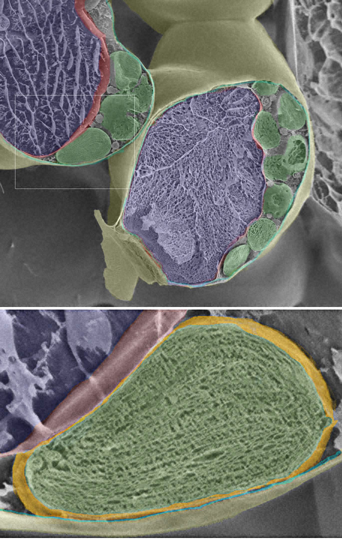
Leaf chloroplast - ru High-resolution SEM image of the sponge paremchyma cells and the chloroplast © with labels or without labels (172 KB). Printable version of this page in Word or pdf format. TEM view of a single chloroplast and details Chloroplast of tobacco: 1 = cell wall 2 = cytoplasm 3 = vacuole 4 = chloroplast envelope (2 membranes) 5 = tonoplast
A brief history of how microscopic studies led to the elucidation of ... Oct 5, 2020 ... The development of the transmission electron microscope ... This claim was supported by images of chloroplasts that were devoid of internal ...
Arabidopsis CURVATURE THYLAKOID1 Proteins Modify Thylakoid Architecture ... (A) Scanning electron microscopy images of envelope-free chloroplasts from wild-type , CURT1A-HA curt1a-2, and oeCURT1A-cmyc curt1a-2 plants, following immunogold labeling with antibodies raised against CURT1B, cytochrome f, HA, and cmyc. Bars = 500 nm. (B) Quantification of the distribution of immunogold-labeled CURT1 proteins.
Regulation of Chlorophagy during Photoinhibition and Senescence ... TEM images show that vacuolar chloroplasts are partially fragmented, supporting the notion that vacuolar chloroplasts are in the process of being digested ( Fig. 1C ). Such observations led to the discovery of chlorophagy, a process by which whole photodamaged chloroplasts are transported into the central vacuole ( Fig. 1D; Izumi et al. 2017 ).
Chloroplast Photos and Premium High Res Pictures - Getty Images chloroplast structure 4,517 Chloroplast Premium High Res Photos Browse 4,517 chloroplast stock photos and images available, or search for chloroplast micrograph or chloroplast structure to find more great stock photos and pictures. NEXT
TEM Chloroplast Labeling Diagram | Quizlet Start studying TEM Chloroplast Labeling. Learn vocabulary, terms, and more with flashcards, games, and other study tools. Home Browse. Browse. Languages. English French German Latin Spanish View all. Science. Biology Chemistry Earth Science Physics Space Science View all. Arts and Humanities.
Assignment 6, page 2 - North Carolina State University Now, it is time to view a real chloroplast by transmission electron microscopy. Study this transmission electron micrograph of a spinach leaf cell, locate a chloroplast and capture the image for labeling.The micrograph is displayed as if using a "virtual electron microscope", so you will need to magnify the image and move to a region that contains the clearest view of chloroplast internal ...
Three‐dimensional ultrastructure of chloroplast pockets formed under ... Traced and three-dimensional images of a chloroplast with an open type pocket. (a) Part of the traced serial transmission electron microscopy images of a chloroplast with an open type pocket. The number in each photograph indicates the order of serial sections. Green and purple indicate a chloroplast and lipid body, respectively.
chloroplast | Definition, Function, Structure, Location, & Diagram A chloroplast is a type of plastid (a saclike organelle with a double membrane) that serves as the site of photosynthesis, the process by which energy from the Sun is converted into chemical energy for growth. Chloroplasts contain the pigment chlorophyll to absorb light energy.
Chloroplast Micrograph Stock Photos and Images - Alamy Chloroplast, TEM ID: HRJJ1H (RM) Volvox green algae, light micrograph ID: 2FMBTWE (RF) Microscopic view of moss leaf (Plagiomnium affine). Brightfield illumination. ID: 2C85ED4 (RF) Chloroplast, TEM ID: HRJJ1G (RM) Plant stem, light micrograph. ID: 2DAMBD7 (RF) Plant cells under the microscope, showing chlorophyll content ID: 2H9B800 (RM)
Transmission electron microscopy (TEM) images of chloroplasts ... Download scientific diagram | Transmission electron microscopy (TEM) images of chloroplasts from the primary leaf of control (a), NO2-treated (b-d) and ...
1,651 Chloroplast Stock Photos - Dreamstime 1,651 Chloroplast Photos - Free & Royalty-Free Stock Photos from Dreamstime 1,651 Chloroplast Stock Photos Most relevant Best selling Latest uploads Within Results People Pricing License Media Properties More Safe Search plant chloroplast structure chebula terminalia animal cells plant bacteria ulva lactuca cell microscope plant cell plant
Tem Chloroplast Stock Photos and Images - Alamy Find the perfect tem chloroplast stock photo. Huge collection, amazing choice, 100+ million high quality, affordable RF and RM images. No need to register, buy now! ... Search Results for Tem Chloroplast Stock Photos and Images (54) Page 1 of 1. 1. 292,070,161 stock photos, vectors and videos. Buying from Alamy. Licenses and pricing; Browse by ...
Native architecture of the Chlamydomonas chloroplast revealed by in ... Chloroplast function is orchestrated by the organelle's intricate architecture. By combining cryo-focused ion beam milling of vitreous Chlamydomonas cells with cryo-electron tomography, we acquired three-dimensional structures of the chloroplast in its native state within the cell. Chloroplast envelope inner membrane invaginations were frequently found in close association with thylakoid tips ...
PDF Plant Anatomy: Images and diagrams to explain concepts 1.6 CHLOROPLASTS Chloroplasts are large organelles and their function is the formation and storage of carbohydrates from photosynthesis. The chloroplast is bounded by a double membrane. The matrix of the chloroplast is known as the stroma. Also inside the chloroplast are separate internal membranes that form lamellae or rounded tongue-like
Structure of Chloroplast (With Diagram) | Botany - Biology Discussion This will help you to draw the structure and diagram of chloroplast. 1. Chloroplasts (Figs. 295-296), responsible for the photosynthesis of the plants, are the characteristic features of the cells of green plants. ADVERTISEMENTS: 2. Around the chloroplast is present a double membrane envelope. 3. Each membrane of chloroplasst is 35 to 50 Å thick.
Electron Microscopy Images - Dartmouth Sep 10, 2021 ... Transmission electron microscope image of a thin section cut through the bronchiolar epithelium of the lung(mouse), which consists of ...














Post a Comment for "41 tem image of chloroplast labeled"