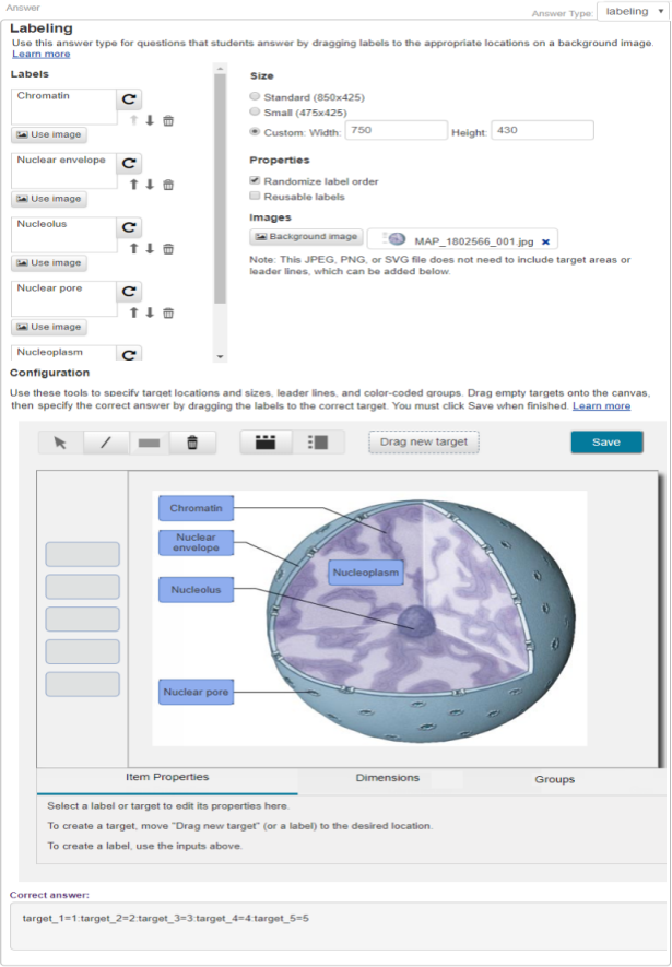
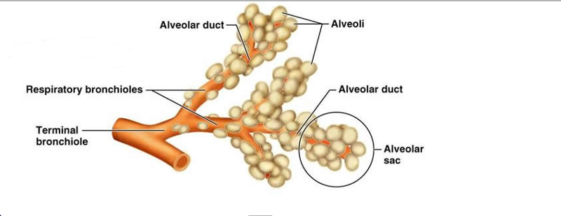
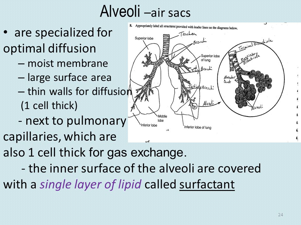
40 appropriately label all structures provided with leader lines on the diagrams below
review sheet exercise 36 Flashcards - Easy Notecards review sheet exercise 36 Flashcards - Easy Notecards. Set Details Share. created 11 years ago by mini. 14,727 views. label all structures provided with leader lines on the diagram below. show more. List View. Show: All Cards 2. PDF Chapter 7 diagrams - Weebly Figure 5—13 is a diagram of the articulated skeleton, Identify all bones or groups of bones by writing the correct labels at the end of the leader lines. Then, select two different colors for the bones of the axial and appendicular skeletons and use them to color in the coding circles and corresponding structures in the diagram.
PDF NS Activities Answers - buckeyevalley.k12.oh.us Created Date: 1/23/2014 12:31:42 PM
:max_bytes(150000):strip_icc()/VennDiagram2-dcf415cf11cf4cd1b03b522a984d9516.png)
Appropriately label all structures provided with leader lines on the diagrams below
PDF OpenALG OpenALG PDF Plainfield East High School Subject: Image Created Date: 10/6/2017 12:19:06 PM Solved 8. Appropriately label all structures provided with - Chegg Appropriately label all structures provided with leader lines on the diagrams below. DURA . This problem has been solved! See the answer See the answer See the answer done loading. Can someone label THIS diagram? Please and thank you in advance! :) Show transcribed image text Expert Answer.
Appropriately label all structures provided with leader lines on the diagrams below. PDF The Cell: Anatomy and Division - Holly H. Nash-Rule, PhD In the following diagram, label all parts provided with a leader line. Nuclear envelope Nucleus Ribosomes Mitochondrion Peroxisome Golgi apparatus Rough endoplasmic reticulum Nuclear pore Nucleolus Cytosol Lysosome Centrioles Microtubule Intermediate filaments Microvilli Smooth endoplasmic reticulum Differences and Similarities in Cell Structure 5. PDF The Skeletal System Answer Key - Weebly BONES—AN OVERVIEW 1. Classify each of the following terms as a projection (P) or a depression or opening (D). Enter the appropriate letter in the answer blanks. 1. Condyle 2. Crest 3. Fissure 4. Foramen 5. Head 6. Meatus 7. Ramus 8. Spine 9. Tuberosity 2. Group each of the following bones into one of the four major bone cate- gories. BIO 202 EX 36.pdf - -b,8 - REVIEW SHEET Anatomy of the... Complete the labeling of the diagram of the upper respiratory structures (sagittal section). \p iot go' P} ... Ll" /' Appropriately label all structures prpvided with leader lines on the diagrams below. JrrroL [,.,;ri^ b*nrl,,i 5:tu' 6 ... The Axial Skeleton - City University of New York A colored dot at the end of a leader line indicates a bone. Leader lines without a colored dot indicate bone markings. Note that vomer, sphenoid bone, and zygomatic bone will each be labeled twice. Key: 1. alvelolar processes 2. carotid canal 3. ethmoid bone (perpendicular plate) 4. external occipital protuberance 5. foramen lacerum / 6.
PDF Human Anatomy & Physiology Laboratory Manual fluids or those provided by the instructor. In many cases, suitable alternatives have been suggested. All reusable glassware and plasticware should be soaked in 10% bleach solution for 2 hours and then washed with laboratory detergent and autoclaved if possible. Disposable items should be placed in an autoclave bag for 15 BI217 Sec.750 Spring 2017: Exercise 36 Flashcards | Quizlet Appropriately label all structures provided with leader lines on the diagrams below. ... 9. Trace a molecule of oxygen from the nostrils to the pulmonary capillaries of the lungs: Nostrils → NOSTRILS -> NASAL CAVITY -> PHARYNX -> LARYNX -> TRACHEA -? A & P II Lab Practical 2 Review Flashcards | Quizlet Identify each; on the lines to the sides, not the structural detail that enable you to make these identifications: Artery - open, circular lumen (always patent) - Thick tunica media (smooth muscle) - elatsin Identify each; on the lines to the sides, not the structural detail that enable you to make these identifications: Vein - Semi-collapsed lumen 8 Appropriately label all structures provided with leader lines on the ... See Page 1. 8 Appropriately label all structures provided with leader lines on the diagrams below. 9 .9 Trace a molecule of oxygen from the nostrils to the pulmonary capillaries of the lungs: Nostrils → 10 Match the terms in column B to the descriptions in column A. Column A0 1. connects the larynx to the main bronchi 0 2. includes terminal ...
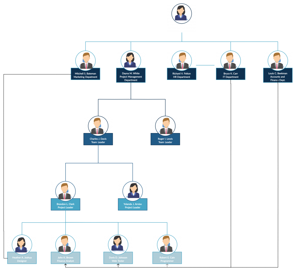
ReaderUi ReaderUi The Structure of DNA - University of Arizona Each nucleotide is itself make of three subunits: A five carbon sugar called deoxyribose (Labeled S) A phosphate group (a phosphorous atom surrounded by four oxygen atoms.) (Labeled P) And one of four nitrogen-containing molecules called nucleotides . (Labeled A, T, C, or G) PDF Document2 - Gore's Anatomy & Physiology 3. each of the eye muscles indicated by leader lines an l. 'gure Color code and color each muscle a different color Then. in the blanks below, indicate (he mo.' ement caused by muscle rectus Supenor oblique 6 Inferior oblique Optic nerve Figure 8-1 Three eye structures contribute to tòrmation oi tears Ad cycbail the: 'Able, name stmcture and 5 Best Organizational Structure Examples (For Any Business) Project D. Marketing Team (D) Operations Team (D) Finance Team (D) HR Team (D) This hybrid organizational structure example tries to combine a functional organizational structure with a matrix-based one. In this instance, the business is also project-based, but the team follows a functional structure.
unioncc.instructure.com You are being redirected.
Draw a labelled diagram of reflex arc and explain reflex action. Solution. The reflex arc describes the pathway in which the nerve impulse is carried and the response is generated and shown by the effector organ. 1. The receptor is present in the receptor organ. 2. The sensory neuron conducts the nerve impulses towards the central nervous system (CNS). The CNS is comprised of the brain and the spinal cord. 3.
Anatomy And Physiology Coloring Workbook A Complete Study ... - Calameo 88 Anatomy & Physiology Coloring Workbook 21. Identify the bones in Figure 5 -9 by labeling the leader lines identified as A, B, and C. Color the bones different colors. Using the following terms, com plete the illustration by labeling all bone markings provided with leader lines.
PDF Chapter 3 Review Materials Key - wtps.org Name the cytoskeletal element (microtubules, microfilaments, or intermediate filaments) described by each of the following phrases. m CEO {IR 1. give the cell its shape 2. resist tension placed on a cell I C/Lc 3. radiate from the cell center 4. interact with myosin to produce contractile force are the most stable 6. have the thickest diameter 7.
UML Diagram Types | Learn About All 14 Types of UML Diagrams A component diagram displays the structural relationship of components of a software system. These are mostly used when working with complex systems with many components. Components communicate with each other using interfaces. The interfaces are linked using connectors. The image below shows a component diagram.
PDF Document1 - Gore's Anatomy & Physiology Identify the bones and bone markings indicated by leader lines on the figure. Select different colors tor the structures listed below and use them to color the coding circles and corresponding structures in the figure. Also, label the dashed line shaw- ing rhe dimensions of the true pelvis and that showing the diameter of the false pelvis.
Solved 8. Appropriately label all structures provided with - Chegg Expert Answer 100% (13 ratings) The larynx is a tough and flexible segment of the respiratory tract connecting the pharynx to the trachea of … View the full answer Transcribed image text: 8. Appropriately label all structures provided with leader lines on the model shown below. Previous question Next question
DOC Respiratory System 23 - PC\|MAC Figure 23-1 is a sagittal view of the upper respiratory structures. First, correctly identify all structures provided with leader lines on the figure. Then select different colors for the structures listed below and use them to color in the coding circles and the corresponding structures on the figure. nasal cavity larynx pharynx trachea
Structure and Functions of Human Eye with labelled Diagram - BYJUS Conjunctiva: It lines the sclera and is made up of stratified squamous epithelium. It keeps our eyes moist and clear and provides lubrication by secreting mucus and tears. Cornea: It is the transparent, anterior or front part of our eye, which covers the pupil and the iris. The main function is to refract the light along with the lens.
PDF Home - Buckeye Valley Home - Buckeye Valley
Solved > 20.Label the tissue types illustrated here and on:1609441 ... View Solution 1.Complete the following statements by writing the appropriate word or phrase on the correspondingly numbered blank: The two basic tissues of which the skin is composed... 3.Using the key choices, choose all responses that apply to the following descriptions. Some terms are used more than once.
Solved 8. Appropriately label all structures provided with - Chegg Appropriately label all structures provided with leader lines on the diagrams below. DURA . This problem has been solved! See the answer See the answer See the answer done loading. Can someone label THIS diagram? Please and thank you in advance! :) Show transcribed image text Expert Answer.
PDF Plainfield East High School Subject: Image Created Date: 10/6/2017 12:19:06 PM
PDF OpenALG OpenALG


















Post a Comment for "40 appropriately label all structures provided with leader lines on the diagrams below"