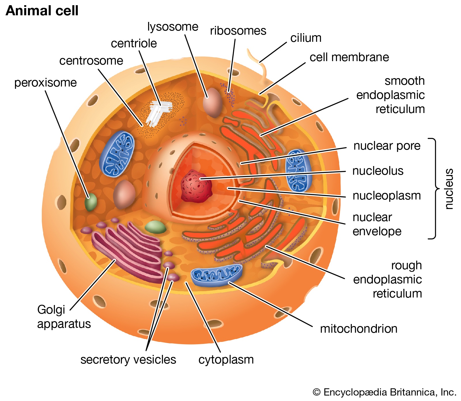
41 identify the structures in the cell pictured on the right. label a label b label c label d label e
Science S1 Flashcards | Quizlet Study with Quizlet and memorize flashcards containing terms like In which order did the events forming our solar system occur? The solar nebula became hot and dense pulling in more gas.This flattened into a rotating disk. It spun faster and faster, forming the Sun. Gas was pulled toward the center, forming the Sun. Gas flattened into a rotating disk and became hot and dense, forming a solar ... Prokaryotic Cells: Structure, Function, and Definition - ThoughtCo Prokaryotes are single-celled organisms that are the earliest and most primitive forms of life on earth. As organized in the Three Domain System, prokaryotes include bacteria and archaeans. Some prokaryotes, such as cyanobacteria, are photosynthetic organisms and are capable of photosynthesis . Many prokaryotes are extremophiles and can live ...
Identify the structures in the cell pictured on the right. - Brainly Find an answer to your question Identify the structures in the cell pictured on the right. dipaleep3794 dipaleep3794 15.01.2019 Biology Secondary School answered Identify the structures in the cell pictured on the right. 2 See answers Advertisement

Identify the structures in the cell pictured on the right. label a label b label c label d label e
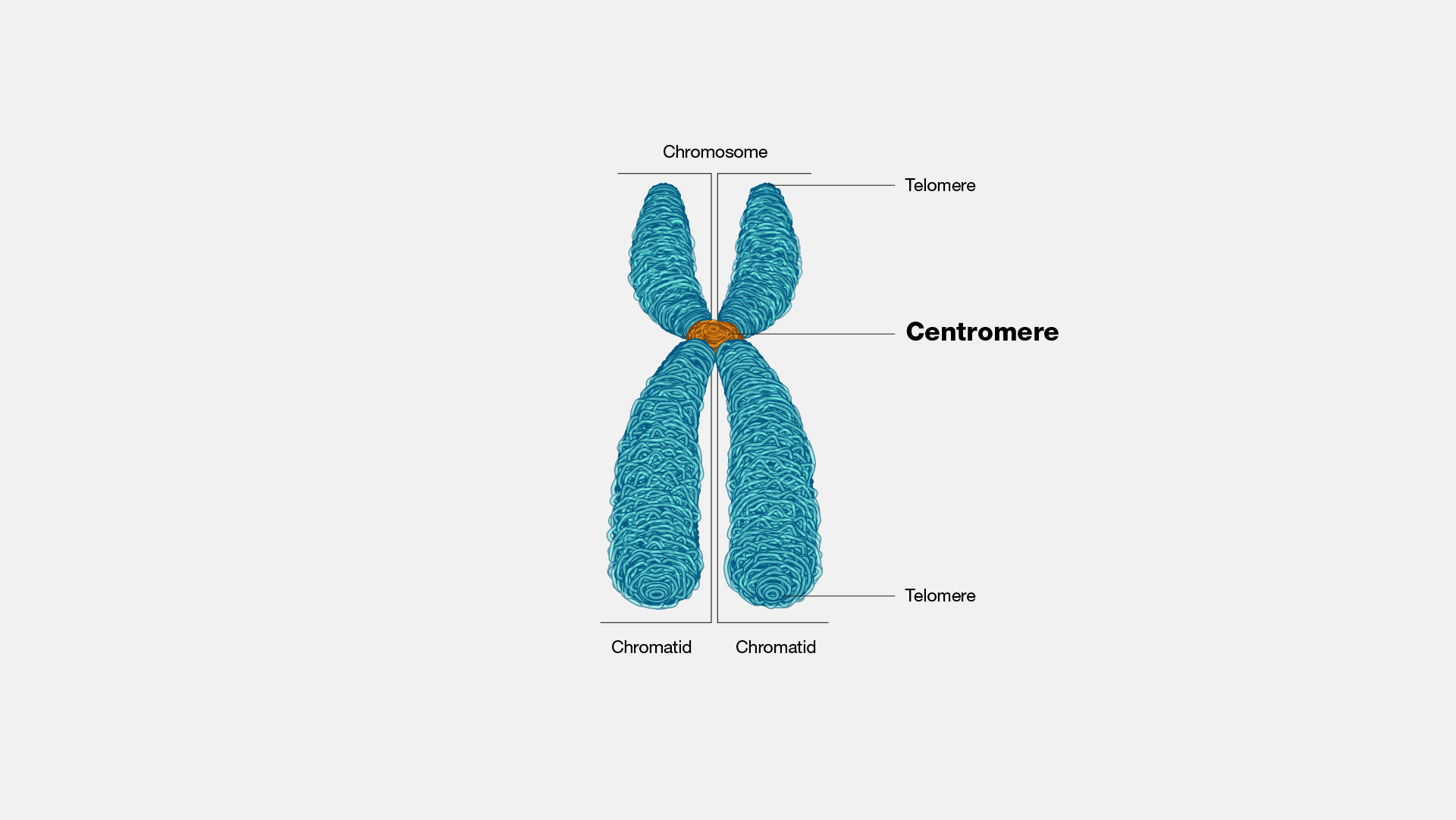
Note Making Class 11 CBSE Format, Examples – Learn Cram Aug 25, 2020 · Main idea: Identify main idea from TOPIC SENTENCE (if there is one) or use BASIC SIGNAL WORDS. C. Identify Supporting Details D. Disregard unimportant information E. Analyse redundant information F. Simplify, categorise, and label important information. Note: Make sure that it does not exceed 1/3 of the length of the original text. Chromosome Structure (Labeling) - The Biology Corner This simple worksheet shows a diagram of a chromosome and where it is located in the nucleus of the cell. Students use a word bank to label the chromatid, centromere, chromosomes, cell membrane, DNA, and nucleus. This worksheet was created for introductory biology for students to practice labeling the parts of a chromosome. Grade Level: 6-12 Chapter 17, Problem 44P | bartleby A cable with a linear density of =0.2 kg/m is hung from telephone poles. The tension in the cable is 500.00 N. The distance between poles is 20 meters.
Identify the structures in the cell pictured on the right. label a label b label c label d label e. Label the structures Flashcards | Quizlet Start studying Label the structures. Learn vocabulary, terms, and more with flashcards, games, and other study tools. Identify the structures in the cell pictured on the right The cell structures are labelled:. A - Nucleus. B - Cytoplasm. C - Ribosomes. D - DNA. E - Cell Membrane/Plasma Membrane . The following are the cell organelles/structures of a eukaryotic cell that is labelled in the image:. Label A - Nucleus: This is as seen in the image is bounded by an outer membrane. It contains the genome that is responsible for replication and transcription of DNA. Mastering Biology Chapter 4 Flashcards | Quizlet Drag the labels onto the diagram to identify the structures of an animal cell. a. cytoskeleton b. ribosomes c. nucleus d. smooth endoplasmic reticulum (ER) e. cytosol f. Golgi apparatus ... Drag the correct label under each cell structure to identify whether it is found only in animal cells, only in plant cells, or in both types of cells. ... Identify the structures in the cell pictured on the right. Label A ... Identify the structures in the cell pictured on the right. Label A Label B Label C Label D Label E Get the answers you need, now! haleighhight0 haleighhight0 05/26/2022 Biology College answered Identify the structures in the cell pictured on the right. Label A Label B
Science 6th Grade part 2 Flashcards | Quizlet A scanning electron microscope is a microscope that sweeps a beam of electrons over the surface of an object to create a three-dimensional image of the object. Only surface of the specimen. Magnification ability of 60,000 times without clarity loss; 500,000 times with losing clarity. Scanning Tunneling Microscope (STM) Using the drop-down menus, identify the structures common to all cells ... Correct answers: 3 question: Using the drop-down menus, identify the structures common to all cells. Label A Label B Label C Label D Answered: Label the various leukocytes. Drag the… | bartleby A pair of structures that connect the axial skeleton to the upper limbs is known as the pectoral girdle. It forms articulations, or joints, with the upper limbs, each consisting of a clavicle and scapula. The human muscular system disorder influences the most important part of the human body- the muscle. PDF Cell City Worksheet Answer Key - Johns Hopkins University 2. The cell membrane is a thin, flexible envelope that surrounds the cell. It allows the cell to change shape and controls what goes into and out of the cell a. What company or place does the cell membrane resemble in a Cell City? City Limits b. Why do you think so? The cell membrane controls what goes into and out of the cell as the city limits
Labeled Neuron Diagram | Science Trends Neurons are a type of cell and are the fundamental constituents of the nervous system and brain. Neurons take in stimuli and convert them to electrical and chemical signals that are sent to our brain. There are 3 major kinds of neurons in the spinal cord: sensory, motor, and interneurons. Neurons communicate vie electrical signals produced by ... Identify the structures in the cell pictured on the right. Label A ... Identify the structures in the cell pictured on the right. Label A Label B Label C Label D Label E Get the answers you need, now! alyssamarie2719 alyssamarie2719 04/11/2020 Biology Middle School answered Identify the structures in the cell pictured on the right. Label A Label B Biochemistry PDF | PDF | Cell (Biology) | Biochemistry - Scribd Cytoskeleton The cytoskeleton acts to organize and maintain the cell's shape; anchors organelles in place; helps during endocytosis, the uptake of external materials by a cell, and cytokinesis, the separation of daughter cells after cell division; and moves parts of the cell in processes of growth and mobility. The eukaryotic cytoskeleton is ... AP1 Lab Manual_Answers - Anatomy and Physiology Lab Manual ... - StuDocu Introduction to Christian Thought (D) (THEO 104) chemistry (BLAW 2001) University Physics Ii (PHYS 2074) Technology Elective (IT1500) Infectious Diseases (CPH 2212) Bachelor of Secondary Education Major in Filipino (BSED 2000, FIL 201) Professional Career Development Seminar (NUR 4828) Early Childhood Foundations and the Teaching Profession ...
Solved Label each phase of the cell cycle in the figure - Chegg Expert Answer. 99% (89 ratings) Transcribed image text: Label each phase of the cell cycle in the figure below with the appropriate name or description. mitosis and cytokinesis Go phase G1 phase DNA replication last stage of interphase.
eHarcourtSchool.com has been retired - Houghton Mifflin Harcourt Connected Teaching and Learning. Connected Teaching and Learning from HMH brings together on-demand professional development, students' assessment data, and relevant practice and instruction.
Cell Organelles- Definition, Structure, Functions, Diagram Cell Organelles Definition. Cell organelles are specialized entities present inside a particular type of cell that performs a specific function. There are various cell organelles, out of which, some are common in most types of cells like cell membranes, nucleus, and cytoplasm. However, some organelles are specific to one particular type of cell ...
Identify the organelles in the cell to the right. A B C D E F The ... C centriole D Intracellular space E extracellular space F Endoplasmic reticulum. Explanation: mitochondria cellular organism which has its own DNA and it is present in both plants and animal cell.The mitochondria is said to the powerhouse of the cell because it helps in the aerobic respiration and generation of large number of ATP by the ...
Cell Organelles - Label | Biology Quiz - Quizizz 30 Questions Show answers Question 1 30 seconds Q. Label A answer choices Cell wall cytoplasm nucleus cell membrane Question 2 300 seconds Q. Name structure #5. answer choices rough ER nucleus vesicle cytoplasm Question 3 300 seconds Q. Name structure #6. answer choices rough ER Golgi body Smooth ER cytoplasm Question 4 30 seconds Q.
Plant Cells Vs. Animal Cells (With Diagrams) - Owlcation The vacuole has an important structural function, as well. When filled with water, the vacuole exerts internal pressure against the cell wall, which helps keep the cell rigid. A plant that is wilting has vacuoles that are no longer filled with water. While animal cells do not have a cell wall, chloroplasts, or a large vacuole, they do have one ...
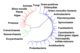
Archaea - Wikipedia In Euryarchaeota the cell division protein FtsZ, which forms a contracting ring around the cell, and the components of the septum that is constructed across the center of the cell, are similar to their bacterial equivalents. In cren-and thaumarchaea, the cell division machinery Cdv fulfills a similar role. This machinery is related to the ...
Identify the structures in the cell pictured on the right. Label A ... 🔴 Answer: 3 🔴 on a question Identify the structures in the cell pictured on the right. Label A Label B Label C Label D Label E nucleus cytoplasm DNA cell membrane ribosomes - the answers to ihomeworkhelpers.com. Subject. English; History; Mathematics; Biology; Spanish; Chemistry;
Identifying eukaryotic cell structures (practice) | Khan Academy How well can you identify eukaryotic cell structures? If you're seeing this message, it means we're having trouble loading external resources on our website. ... Practice: Identifying eukaryotic cell structures. This is the currently selected item. Practice: Eukaryotic cell structures. Next lesson. Prokaryotes and eukaryotes.
(PDF) INTERCHANGE 5ta EDICION - Academia.edu Enter the email address you signed up with and we'll email you a reset link.
Prokaryotic and Eukaryotic cells Flashcards | Quizlet what are the differences in the internal structures of the cells pictured to the right there are fewer structures in the cell on the top and the structures in the cells are similar Which cell structures are seen in all cell types? DNA,cytoplasm,ribosomes,cell membrane
Cell Structure and Organelle Labeling Flashcards | Quizlet identify the type of cell. cell. smallest living unit of life. cell theory. all living things are made of cells, cells come from other cells, cell is the smallest living thing. ... Cell Structures Diagrams. 16 terms. Berlyn_Seitz. Cell Organelle Labeling. 15 terms. PhillipsBio. OTHER SETS BY THIS CREATOR. Zoology - Animal Characteristics. 32 terms.
Cell: Structure and Functions (With Diagram) - Biology Discussion Prokaryotes are the simplest cells without a nucleus and cell organelles. 2. Prokaryotic cells are the smallest cells (1-10 μm). 3. Unicellular and earliest to evolve (~4 billion years ago), still available. 4. The cell wall is rigid. ADVERTISEMENTS: 5. These cells reproduce asexually. 6. They include bacteria and archaea. 7.
Undergraduate | College of Veterinary Medicine | Washington State ... Undergraduate Degree Programs. Whether you go on to graduate school or start a career, our undergraduate degree programs prepare you to be a leader in human and animal science, health, and medicine. Biochemistry, genetics and cell biology, microbiology, and neuroscience majors provide the pre-health education to succeed in dentistry, medicine ...
A Labeled Diagram of the Plant Cell and Functions of its Organelles The cell membrane is a thin layer made up of proteins, lipids, and fats. It forms a protective wall around the organelles contained within the cell. It is selectively permeable and thus, regulates the transportation of materials needed for the survival of the organelles of the cell. Function: Protects the cell from its surroundings. Cell Wall
Chapter 17, Problem 44P | bartleby A cable with a linear density of =0.2 kg/m is hung from telephone poles. The tension in the cable is 500.00 N. The distance between poles is 20 meters.
Chromosome Structure (Labeling) - The Biology Corner This simple worksheet shows a diagram of a chromosome and where it is located in the nucleus of the cell. Students use a word bank to label the chromatid, centromere, chromosomes, cell membrane, DNA, and nucleus. This worksheet was created for introductory biology for students to practice labeling the parts of a chromosome. Grade Level: 6-12
Note Making Class 11 CBSE Format, Examples – Learn Cram Aug 25, 2020 · Main idea: Identify main idea from TOPIC SENTENCE (if there is one) or use BASIC SIGNAL WORDS. C. Identify Supporting Details D. Disregard unimportant information E. Analyse redundant information F. Simplify, categorise, and label important information. Note: Make sure that it does not exceed 1/3 of the length of the original text.





![Expert Answer] Identify the structures in the cell pictured ...](https://us-static.z-dn.net/files/d41/0f74ee3e55de914eb4ce0b4d9f7f3f9e.png)








Post a Comment for "41 identify the structures in the cell pictured on the right. label a label b label c label d label e"