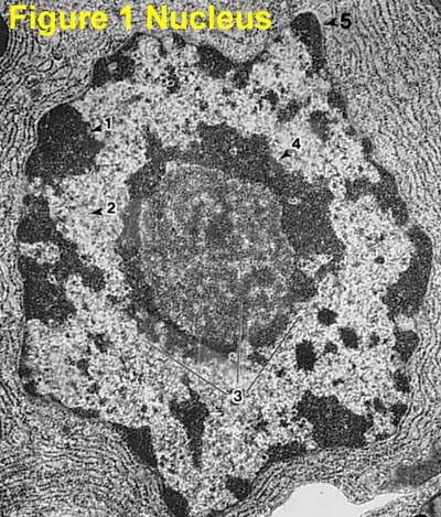
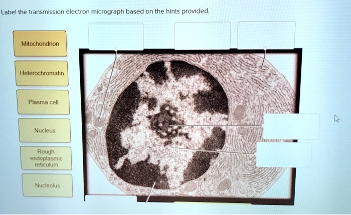
39 label the transmission electron micrograph of the nucleus.
#6 Summary of Cell structure | Biology Notes for A level - Blogger 5. List ten structures you could find in an electron micrograph of an animal cell which would be absent form the cell of a bacterium. 6. Advice on answering question 6: If you are asked to distinguish between two things, it is likely that it is because they have certain things in common and that they may even be confused with each other.In your answer it is helpful where relevant to point out ... Plant Cell Nucleus Electron Micrograph - Dannie Vanlith Below is a collection of electron micrographs with labelled subcellular structures that you should be able to identify. In mammals it's average diameter is about 6 an electron micrograph of a section through an animal cell nucleus (from an insect cell). In flowering plants, this condition occurs in sieve tube elements.74.
Labeling the Cell Flashcards | Quizlet Label the transmission electron micrograph of the nucleus. membrane bound organelles golgi apparatus, mitochondrion, lysosome, peroxisome, rough endoplasmic reticulum nonmembrane bound organelles ribosomes, centrosome, proteasomes cytoskeleton includes microfilaments, intermediate filaments, microtubules Identify the highlighted structures

Label the transmission electron micrograph of the nucleus.
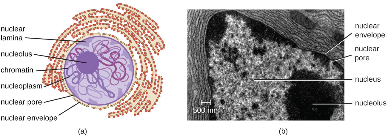
TEM of the Nucleus Unlike fluorescence microscopy, which relies upon the use of fluorescent probes to tag structures, TEM is capable of visualizing the structures themselves. The theoretical resolution of the transmission electron microscope is sufficient to resolve the molecular constituents of the individual nuclear compartments and structures. Cell Nucleus - function, structure, and under a microscope The nucleus is a double-layer membrane organelle. It consists of the nuclear envelope, DNA (chromatin), nucleolus, nucleoplasm, and the nuclear matrix. The main function of the nucleus is to control cell activities and carry genetic information to pass to the next generation. A eukaryotic cell typically has only one nucleus. Improved transmission electron microscopy technique for the study of ... A modification of the technique of Coleman et al for the preparation of single cells in cytologic specimens for electron microscopy (EM) is described. By employing materials in the initial cytologic processing that are useful for EM, such as a paraformaldehyde-glutaraldehyde fixative, lactated Ringe …
Label the transmission electron micrograph of the nucleus.. Fluorescent labeling of resin-embedded sections for correlative ... The arrows in (A) and (B) point towards the same nucleus. (C) Fluorescence micrograph of an area in the mucosa showing staining of nuclei principally in the epithelial layer of obliquely cut crypts of Lieberkühn. The mucous granules of the goblet cells are also labeled (arrowheads). (D) Electron micrograph corresponding to the boxed area in (C). Electron microscopes - Cell structure - Edexcel - BBC Bitesize the transmission electron microscope (TEM) is used to examine thin slices or sections of cells or tissues the scanning electron microscope (SEM) has a large depth of field so can be used to ... Label-free three-dimensional imaging of cell nucleus using third ... The U.S. Department of Energy's Office of Scientific and Technical Information Transmission electron micrograph royalty-free images - Shutterstock Find Transmission electron micrograph stock images in HD and millions of other royalty-free stock photos, illustrations and vectors in the Shutterstock collection. Thousands of new, high-quality pictures added every day.
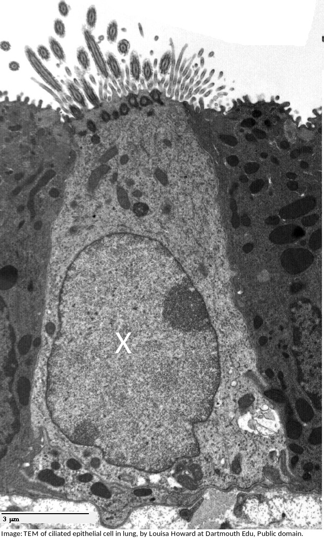
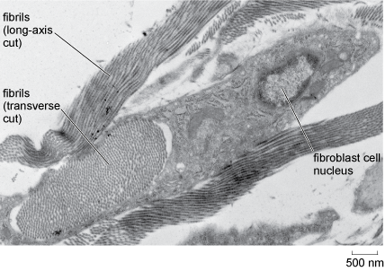
Electron Micrographs of Cell Organelles | Zoology - Biology Discussion This is an electron micrograph of nucleus. (Fig. 17 & 18): (1) Nucleus was discovered by Brown (1831). (2) It is a characteristic entity of almost all eukaryotic cells except mammalian RBCs. (3) The nucleus is generally one but may also be two, four or many. Transmission electron micrograph (TEM) identifying immunogold labeled ... Transmission electron micrograph (TEM) identifying immunogold labeled ESR1 and caveolin-1 proteins in the uterine artery endothelial cells derived from the pregnant state (P-UAEC). (A) IgG control... anatomy 10.png - Label the transmission electron micrograph... anatomy 10.png - Label the transmission electron micrograph of the. anatomy 10.png - Label the transmission electron micrograph... School Utah Valley University; Course Title ZOOL 1090; Uploaded By emileeroylance19. Pages 1 Ratings 67% (3) 2 out of 3 people found this document helpful; PDF Identifying Organelles from an Electron Micrograph Nucleus Chromatin The vacuole in this mature plant cell from a leaf is large, and occupies about 80% of the cell volume The photograph shown below details chloroplast structure as viewed with a transmission electron microscope Courtesy of Dr. Julian Thorpe - EM & FACS Lab, Biological Sciences University Of Sussex
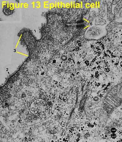
Solved Label the transmission electron micrograph of the - Chegg Expert Answer. 100% (23 ratings) Transcribed image text: Label the transmission electron micrograph of the nucleus. Nuclear envelope Nucleolus Nucleus Heterochromatin Reset Zoom. Light and Electron Microscopy Study of Glycogen Synthase Kinase-3β in ... The subthalamic nucleus is also strongly labeled ( Figure 3C) while adjacent hypothalamic areas present only weakly labeled neurons. In the midbrain, the substantia nigra and ventral tegmental area present GSK3β positive neurons, the substantia nigra pars compacta being the most strongly labeled area ( Figure 3D ). [Solved] Draw and label the ultrastructure of a pancreatic exocrine ... The cell wall, nucleus, vacuoles, mitochondria, endoplasmic reticulum, Golgi apparatus, and ribosomes are easily visible in this transmission electron micrograph. In the above picture, some of the organelles that are clearly recognizable are in the lower central round figure the NUCLEUS. the black dots are the vacuoles. Diagram Of Animal Cell As Seen Under Electron Microscope - Blogger Image:plant cell seen under light microscope the cell as seen under the electron microscope. 80.the diagram below is a drawing of an organelle from a ciliated cell as seen with an electron microscope. Under a high power microscope like the scanning transmission electron microscope, it is possible even to stain and observe the detailed.
Transmission electron micrographs of seminiferous tubules of ...
Bio101 - Ch 6 HW Flashcards | Quizlet -transmission electron microscopy (TEM) to study the movement of organelles within a living cell -scanning electron microscopy (SEM) to study the detailed movements of living cells -cell fractionation to study the function of specific organelles Beginning within the nucleus, the first step leading to the synthesis of a polypeptide is __________.
Solved] FIGURE 5.5 Transmission electron micrographs of ...
Label This Transmission Electron Micrograph - Kaiden Brown Label the transmission electron micrograph of the nucleus. Label the transmission electron micrograph of the nucleus. Transmission electron microscopy (tem) is a microscopy technique in which a beam of electrons is transmitted through a specimen to form an image. Figures label this transmission electron micrograph ( 16, . CIN2003. Ian Roberts.

Cell Micrographs | BioNinja
Looking at the Structure of Cells in the Microscope Determining the detailed structure of the membranes and organelles in cells requires the higher resolution attainable in a transmission electron microscope. Specific macromolecules can be localized with colloidal gold linked to antibodies. Three-dimensional views of the surfaces of cells and tissues are obtained by scanning electron microscopy.

Figure, Transmission Electron Micrograph of Rough Endoplasmic ...
Label This Transmission Electron Micrograph / Microscopy Innovations ... Label the transmission electron micrograph of the nucleus. Transmission electron micrographs of hela cell sections labeled in . Label the transmission electron micrograph of the nucleus. Fluorescence microscopy in combination with tem and an ion beam analysis (iba, which allows the evaluation of the chemical elemental distribution) has allowed .
Ultrastructure of cells 1.2
Label the transmission electron micrograph of the nucleus. - Transtutors Label the transmission electron micrograph of the nucleus. Expert's Answer Solution.pdf Next Previous Q: Q: Q: Q: Q: Copy And Paste 5 Micrographs With Magnifications That Fall Within The Specified Ranges Into The Text Answer Box. Be Sure To Label Your Images With The Appropriate Name And Magnification. Post Them In The Specified Order. 1.
IB Cell Structure Review (1.1-1.2)
Animal Cell Electron Microscope Labelled - Q14 Draw a large diagram of ... Here is an electron micrograph of an animal cell with the labels superimposed: Make your work easier by using a label. Make your work easier by using a label. After this, add another oval shape outside the line you just drew, and this will make the cell membrane to your animal cell. You see that many features are in common.
Unique Characteristics of Eukaryotic Cells | Microbiology ...
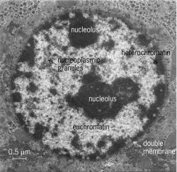
Electron Micrographs - University of Oklahoma Health Sciences Center You should concentrate on the similarities in form that permit identification of the components irrespective of cell type. Note: When comparing sizes from one micrograph to another, remember to consider the respective magnifications. Figure 1 Micrograph of a nucleus. 1. Heterochromatin 2. Euchromatin 3. Nucleolus 4. Nucleolar associated chromatin

Transmission electron-micrograph of the tubal tonsil showing ...
Transmission electron microscopy techniques, Electron microscopy, Otago ... The transmission of unscattered electrons is a function of the specimen thickness and elemental composition. Areas of the specimen that are more electron dense allow fewer transmitted unscattered electrons and appear darker, conversely the thinner areas and those containing lighter elements, permit more transmission and appear lighter.
Electron Micrographs
Neuron under Microscope with Labeled Diagram - AnatomyLearner The nucleus is the spherical or elliptical structure in the neuron containing euchromatic staining (pale staining). Again, the shape of the nucleus of a neuron is generally large because of the little cell body cytoplasm. There is a prominent nucleolus evident in the nucleus of a neuron.

Electron micrographs of SPIO-labeled MSCs. A, Cell nucleus (N ...
Transmission Electron Micrograph of transfected HL-1 cells labeled for ... Transmission Electron Micrograph of transfected HL-1 cells labeled for TMEM43 with immunogold. A and B. Single immunogold labeling experiments used 15 nm gold particles to label GFP. A....
Transmission Electron Microscopy in Cell Biology: sample ...
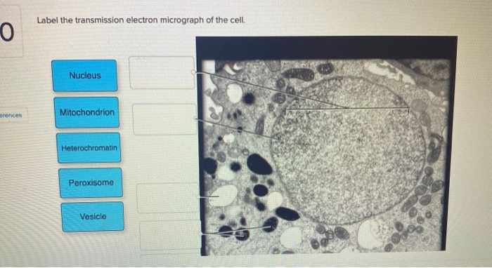
Solved Label the transmission electron micrograph of the - Chegg Question: Label the transmission electron micrograph of the cell. 0 Nucleus rences Mitochondrion Heterochromatin Peroxisome Vesicle ULAR bumit Click and drag each label into the correct category to indicate whether it pertains to the cytoplasm or the plasma membrane.

Structure and Functions of the Dentin-Pulp Complex | Pocket ...
Improved transmission electron microscopy technique for the study of ... A modification of the technique of Coleman et al for the preparation of single cells in cytologic specimens for electron microscopy (EM) is described. By employing materials in the initial cytologic processing that are useful for EM, such as a paraformaldehyde-glutaraldehyde fixative, lactated Ringe …
What is a diagram of a plant and animal cell under an ...
Cell Nucleus - function, structure, and under a microscope The nucleus is a double-layer membrane organelle. It consists of the nuclear envelope, DNA (chromatin), nucleolus, nucleoplasm, and the nuclear matrix. The main function of the nucleus is to control cell activities and carry genetic information to pass to the next generation. A eukaryotic cell typically has only one nucleus.
A tour of the cell: View as single page
TEM of the Nucleus Unlike fluorescence microscopy, which relies upon the use of fluorescent probes to tag structures, TEM is capable of visualizing the structures themselves. The theoretical resolution of the transmission electron microscope is sufficient to resolve the molecular constituents of the individual nuclear compartments and structures.

Identifying the Parts of the Nucleus in an Electron Micrograph
Electron Micrographs

Chromosomes in the nucleus of a cell Stock Photo - Alamy

Transmission electron micrograph of turkey spermatozoa ...

ELECTRON MICROSCOPY — Columbia Nano Initiative

Nucleolus Electron Micrograph

transmission electron micrograph of light cells showing ...
Solved Label the transmission electron micrograph of the ...

Labeling the Cell Flashcards | Quizlet
Histology and cytochemistry of interactions between plants ...

Cell Micrographs | BioNinja
Electron Micrographs
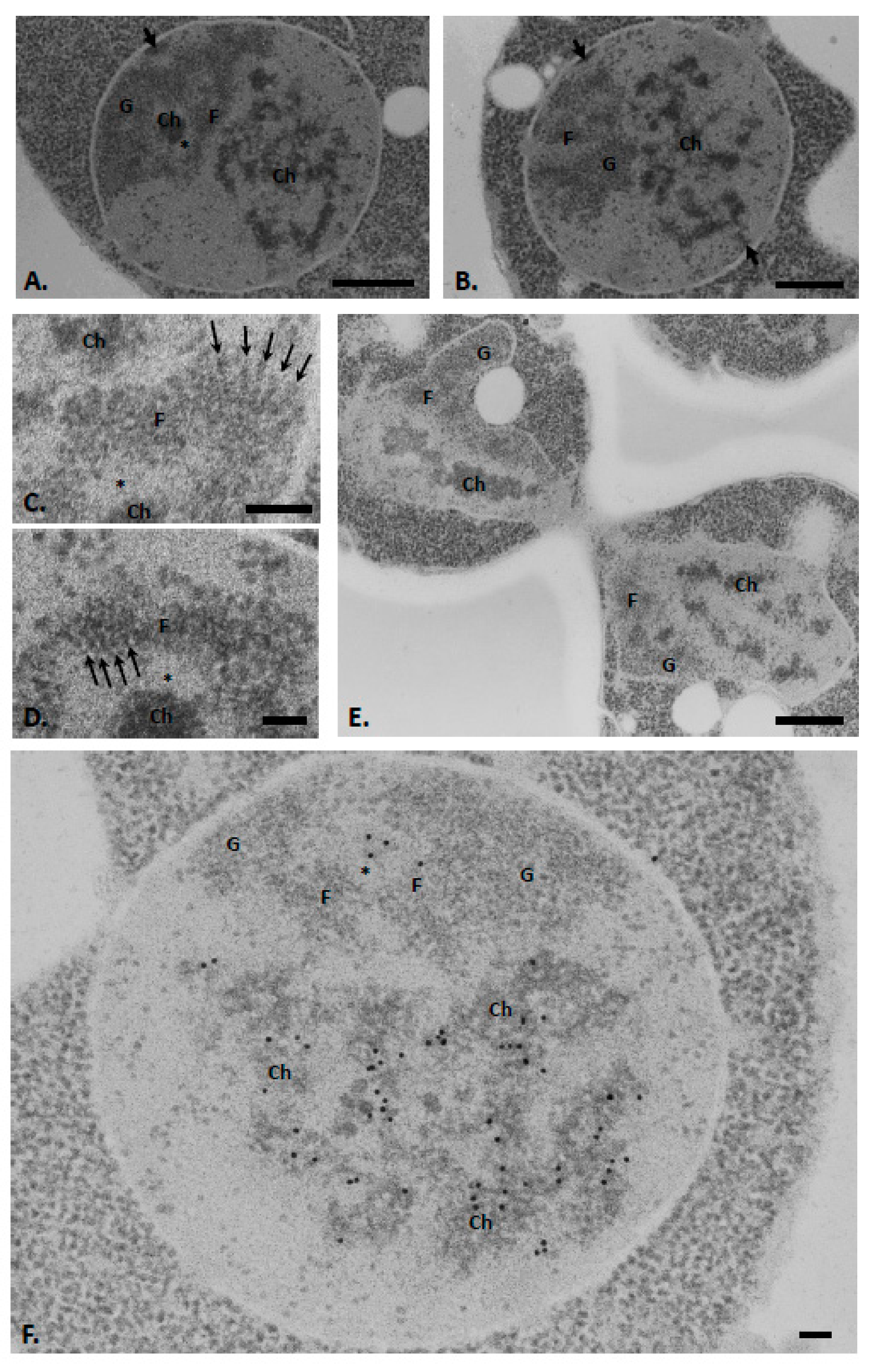
IJMS | Free Full-Text | Visualization of Chromatin in the ...

A Transmission electron micrograph (TEM) of a transverse ...
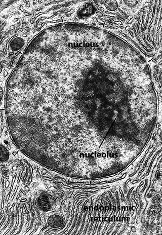
BIOL 230 Lecture Guide - Electron Micrograph of a Nucleus

Cell Structure & Mitosis Visual Lab - ppt download
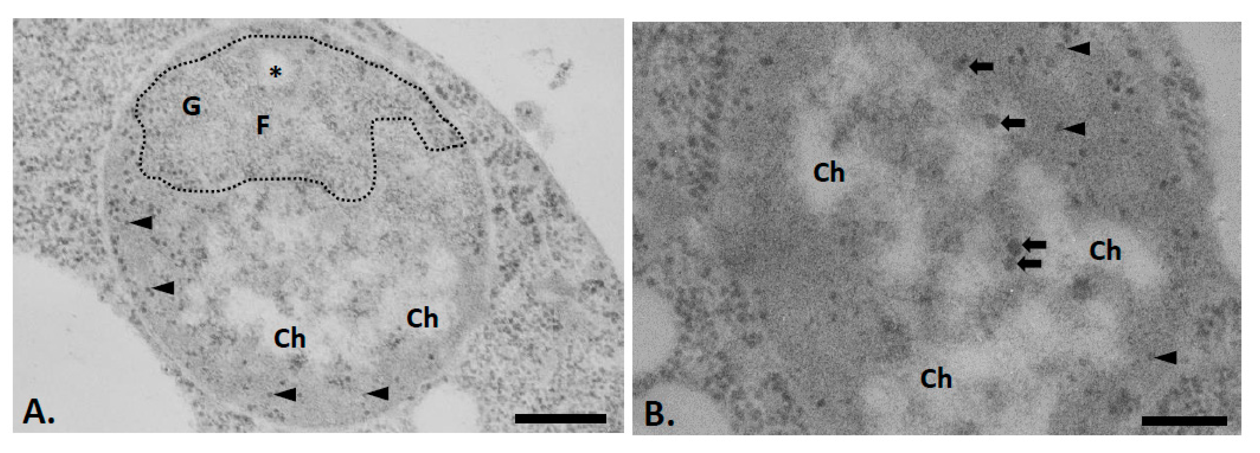
IJMS | Free Full-Text | Visualization of Chromatin in the ...
What is a diagram of a plant and animal cell under an ...

Transmission electron micrograph of an animal cell - Stock ...

PDF) IB Questionbank Test | Ankit Mistry - Academia.edu
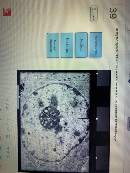
Solved Identify the organelle pictured, then label its ...
Cells (2.1, 2.2, 2.3, 2.4 & 2.5) Flashcards | Quizlet
SOLVED: Label the transmission electron ricrograph based on ...

Transmission electron micrograph of a gold-labelled Lowicryl ...

DP Biology: Ultrastructure of cells quiz 1.2
Post a Comment for "39 label the transmission electron micrograph of the nucleus."