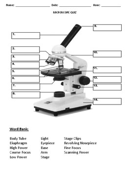
43 label the compound microscope
Quiz Of Microscope Parts And Function the compound microscope uses lenses and light to enlarge the image and is also called an optical or light microscope (versus an electron microscope) liver disease is any abnormal process that affects the liver biology classes often take out a microscope and look at single-celled microbes from pond water change is an essential part of our world, … And Worksheet Parts Microscope Quizlet Use Base: The base of a compound microscope is helps in supporting the microscope and contains the illuminator. Over 10,000 math, reading, grammar and writing, vocabulary, spelling and cursive writing K5 Learning offers free worksheets and inexpensive workbooks for kids in kindergarten to grade 5 Discussion of the brain, and how it works, can be a ...
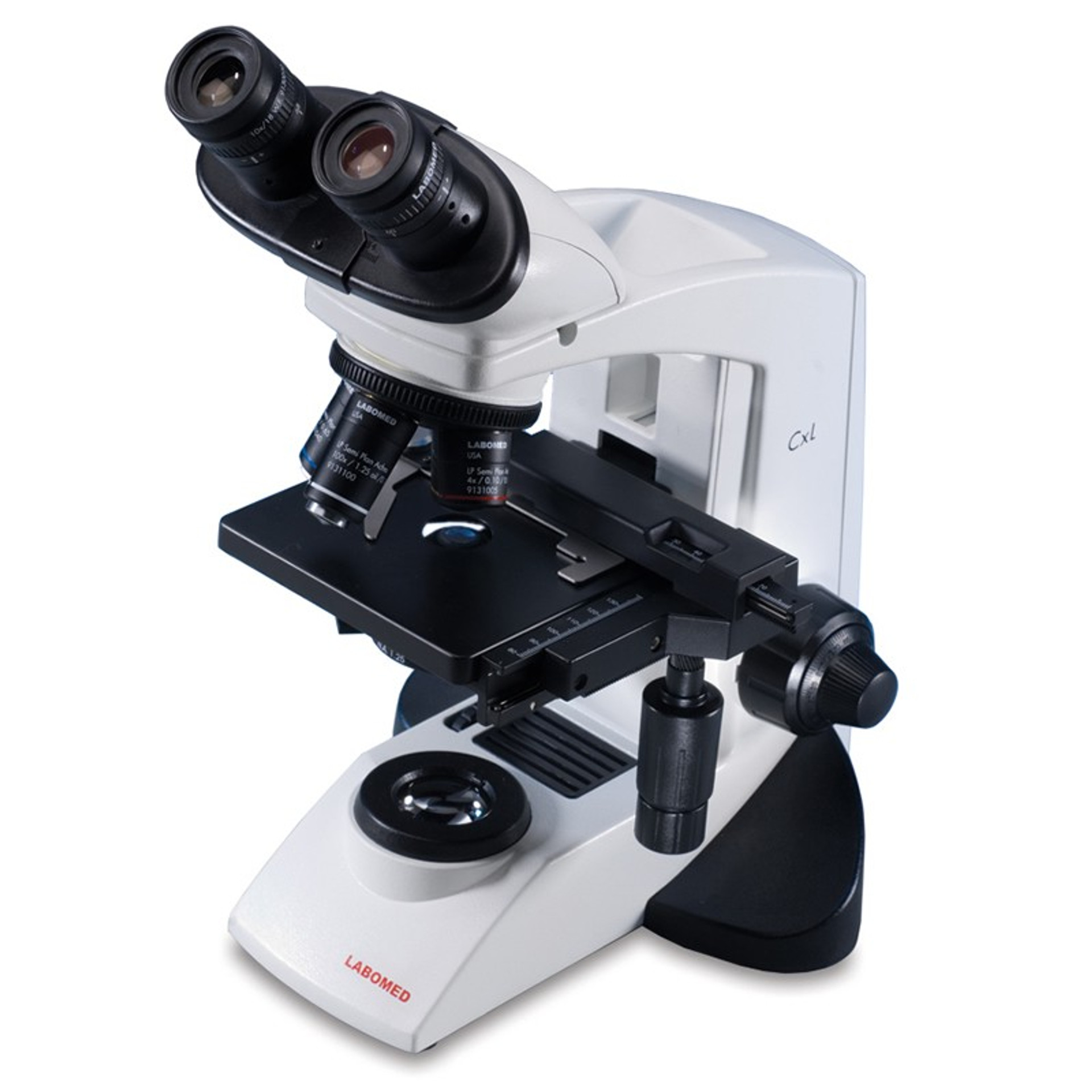
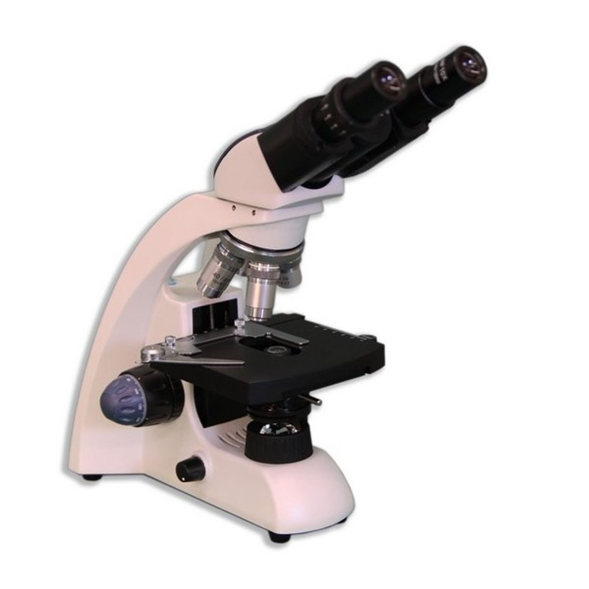
10 Best First Microscope -Reviews & Comparison of 2022 Duo-Scope functions as a compound microscope and stereomicroscope in one unit. Two light sources allow for viewing microscope slides and three-dimensional items such as rocks and leaves. MFL DUO-SCOPE -- Our MFL (My First Lab) Duo Scope is an ideal microscope kit for grades K-12 and comes with individual accessories for young learners.
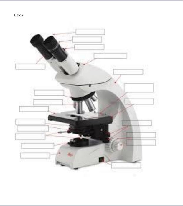
Label the compound microscope
Metaphase - Genome.gov 00:43. Metaphase is a stage during the process of cell division (mitosis or meiosis). Normally, individual chromosomes are spread out in the cell nucleus. During metaphase, the nucleus dissolves and the cell's chromosomes condense and move together, aligning in the center of the dividing cell. At this stage, the chromosomes are ... ECLIPSE Ti2 Series | Inverted Microscopes - Nikon Instruments Inc. The ECLIPSE Ti2 inverted microscope delivers an unparalleled 25mm field of view (FOV) that revolutionizes the way you see. With this incredible FOV, the Ti2 maximizes the sensor area of large-format CMOS cameras without making compromises, and significantly improves data throughput. The Ti2's exceptionally stable, drift-free platform is ... How Microscope Actual Size To Calculate label the parts of the microscope 1 the angle of view of the camera is determined by the ratio of the image size on the film and the focal length draw the letter, exactly the way it looks through the microscope in the following circle, representing the field of view knowing this for each objective lens, you can compare the size of the specimen …
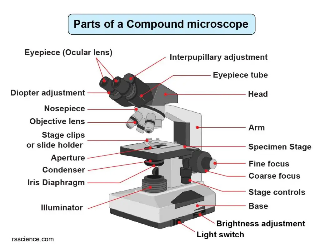
Label the compound microscope. Basic Microscope Diagram - microscope diagram purposegames, images 01 ... Basic Microscope Diagram - 15 images - label the neuron clip art at vector clip art online, microscope diagram fill online printable fillable blank pdffiller, animal anatomy biology4isc, images 01 introduction and terminology basic human anatomy, Light Microscope (Theory) : Cell biology Virtual Lab I : Biotechnology ... The modern compound microscope consists of two lens system, the objective and the ocular or eye piece. The first magnified image obtained with objective lens, is again magnified by the eye piece to give a virtual inverted image. The total magnification the product of the magnifications of two lens systems. Parts of a Microscope R&D 100 Finalists for 2022 are announced Finalists for the 2022 R&D 100 Awards have been announced by R&D World magazine. This renowned worldwide science and innovation competition, celebrating its 60th year, received entries from a dozen different countries and regions. This year's esteemed judging panel included nearly 50 well-respected industry professionals from across the world. The microbiome of a bacterivorous marine choanoflagellate contains a ... The concentrated seawater was labelled with LysoTracker Green DND-26 (Invitrogen), a label that targets acidic food vacuoles of living cells 67, to a final concentration of 25 nM from a working ...
Basic Microscope Diagram - the compound microscope diagram microscopic ... Basic Microscope Diagram - 15 images - microscope 1 light microscope, animal anatomy biology4isc, vehicle body damage inspection diagram, microscope diagram purposegames, A novel approach to develop an animal model for oral submucous fibrosis ... ANE administration by intraoral droplet and intrabuccal injection method exhibits OSF-associated changes in buccal tissues. The hematoxylin and eosin-stained tissue sections under a compound light microscope exhibited OSF-associated changes in the lamina propria and epithelium, as shown in Fig. 2.The intraoral droplet group of ANE administration exhibited a relatively higher level of fibrosis ... Columbia staining jar for microscope coverslips - ProSciTech Columbia staining jar for microscope coverslips. This staining jar can hold up to 4 cover slips measuring 17-23mm wide, and up to 30mm long. Longer coverslips can be accommodated by removing the cap. The jar comes with a white polypropylene screw cap with a PTFE coated polyethylene liner. It is manufactured from Wheaton soda lime glass, which ... proscopehr Power Ball is America's oldest interstate lottery. This lottery was launched in 1992 and is now offered in 45 states, the District of Columbia, Puerto Rico, and the US Virgin Islands. It has the biggest jackpot in history at $1.586 billion, and the winnings can reach six-figures.
Human Biology Lab Online | Lab 2 - Microscopes, Cell Structure and Function Be sure to go through the complete checklist Microscope controls: turn knobs (click and hold on upper or lower portion of knob) throw switches (click and drag) turn dials (click and drag) move levers (click and drag) changes lenses (click and drag on objective housing) select a specimen (click on a slide) Experiment 1 ( Cheah Kheng Keat H8T03B) PDF - Flip eBook Pages 1-9 ... Light compound microscope is a microscope that use light source and has 2 set of lenses, ocular lens and objective lens. We should always hold the microscope with both hands, that is hold the body arm of the microscope with one hand and the base of the microscope with the other hand. We use the oil immersion objective lens to observe CPT® Code 88377 in section: Morphometric analysis, in situ ... CPT® Code 88377 in section: Morphometric analysis, in situ hybridization (quantitative or semi-quantitative) each probe Motility Test (Theory) - Amrita Vishwa Vidyapeetham Virtual Lab When the cover glass is inverted over the well of the slide, the drop hangs from the glass in the hollow concavity of the slide. Since the drop lies within an enclosed glass chamber, drying out occurs very slowly. A ring of Vaseline around the edge of the cover slip keeps the slide from drying out. II) Method for pathogenic microorganisms includes
Compound Microscope Parts, Functions, and Labeled Diagram ...
A Guide to Scanning Transmission Electron Microscopy (STEM) An electron microscope produces images with a resolution that is 1000 times higher than that of an optical microscope. Electron microscopes were constructed as Transmission Electron Microscopes (TEM) and later as Scanning Electron Microscopes (SEM), which provided additional scanning capabilities with magnetic coils, detectors and circuitry.

Compound Microscope- Definition, Labeled Diagram, Principle ...
How To Dissect A Fetal Pig | Carolina.com Compound Microscopes. Popular corded compound microscopes and cordless microscopes for elementary to advanced use. We have the compound microscope you are looking for! Digital Microscopes. Digital microscopes are great for large classroom computer combined instruction. Students can take images, videos, and more. Stereomicroscopes

Compound Microscope – Diagram (Parts labelled), Principle and ...
scheme work biology - Free KCPE Past Papers Organisms in the school compound; Charts on external features of plants and animals; Comprehensive secondary Biology students Bk. 1 page 2-3; Teachers bk. 1 pages 1-4; ... Draw and label the light microscope; Description of a cell; Drawing and labeling the light microscope . Light microscope;

Microscope Components - Science Quiz
Timing of neuron development determines what they can become To do this, the researchers labeled the precursor cells at different times so that all neurons that emerged from these precursors in the following days lit up green under the microscope.

Label the microscope — Science Learning Hub
Labeled Cell Elodea Label in the space below an oblong cell and a spike cell as seen under high power cell membrane, nucleus, and Draw three types of cells (Cheek cell, Red blood cell, Elodea) Place a drop of water on a clean slide Whats people lookup in this blog: Elodea Leaf Under Microscope 400x Labeled Elodea leaf Elodea, also known as Elodea …

Label the microscope — Science Learning Hub
Mr. Jones's Science Class Matter: Atoms and Properties - Open Response Question 3. Force and Motion - Open Response Question 3. Forms of Energy - Open Response Question 1. Forms of Energy - Open Response Question 2. Earth's Structure & Natural Processes - Open Response Question 1.

Photo Compound microscope with labels Image #3850568
Diatom - Wikipedia Diatomaceous earth (diatomite) is a collection of diatom shells found in the earth's crust. They are soft, silica-containing sedimentary rocks which are easily crumbled into a fine powder and typically have a particle size of 10 to 200 μm.

Compound Microscope Olympus, कंपाउंड ...
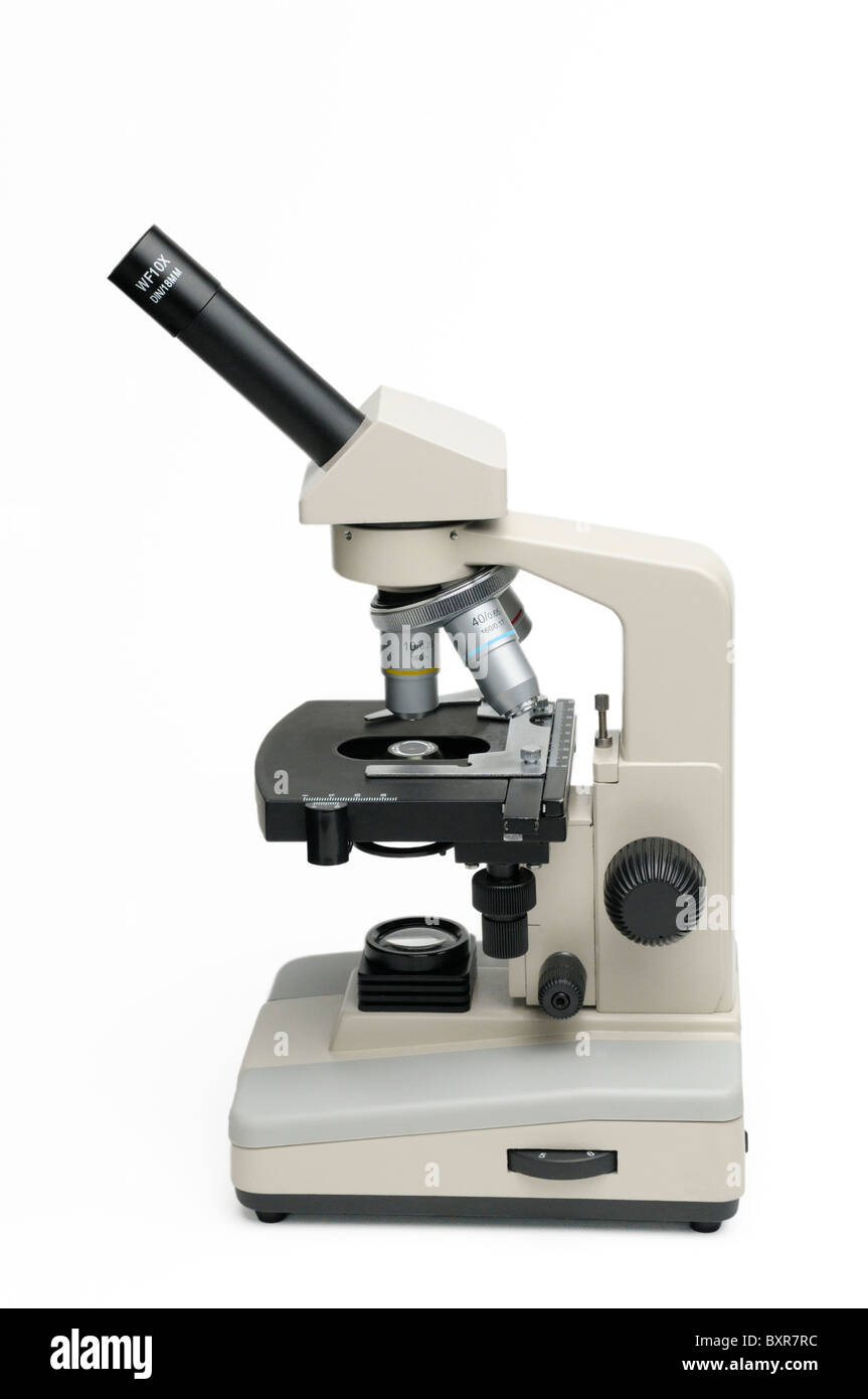
How To Use A Compound Microscope - Bio, Net Worth, Birthday, Height ... How To Use A Compound Microscope? Compound Microscopes. Turn the revolving turret (2) so that the lowest power objective lens (eg. 4x) is clicked into position. Place the microscope slide on the stage (6) and fasten it with the stage clips. Look at the objective lens (3) and the stage from the side and turn the focus knob (4) so the stage moves ...

General Biology | Carlson Stock Art | General biology ...
X-ray Powder Diffraction (XRD) - Techniques What is X-ray Powder Diffraction (XRD) X-ray powder diffraction (XRD) is a rapid analytical technique primarily used for phase identification of a crystalline material and can provide information on unit cell dimensions. The analyzed material is finely ground, homogenized, and average bulk composition is determined.

Microscope labeled diagram
Understanding Your Pathology Report: Melanoma | OncoLink Tumor-Infiltrating Lymphocytes (TILs): TILs look at your immune response to the melanoma. When the pathologist looks at the melanoma under the microscope, they look for the number of lymphocytes (white blood cells) within the lesion. This response, or TILs, is often described as "brisk," "non-brisk," or "absent,".

This is a common compound microscope. Label its parts from A ...
UW SWAP Online Auction Label Printer . Sold $ 55.00. Floor Runner . Sold $ 12.00 Floor Runner . Sold $ 10. ... Inverted Compound Microscope . Sold $ 39.00. Locker . Sold $ 31.00 Lot of 5 Electromagnetic Induction Kit ... Stereozoom Microscope (PARTS / REPAIR) Sold $ 242.00. Rolling Task Chair . Sold $ 30.00. Lot of 2 Rolling Office Chairs ...

Parts of a microscope with functions and labeled diagram
How Microscope Actual Size To Calculate label the parts of the microscope 1 the angle of view of the camera is determined by the ratio of the image size on the film and the focal length draw the letter, exactly the way it looks through the microscope in the following circle, representing the field of view knowing this for each objective lens, you can compare the size of the specimen …

Amscope 40X-1000X Trinocular Biological Compound ...
ECLIPSE Ti2 Series | Inverted Microscopes - Nikon Instruments Inc. The ECLIPSE Ti2 inverted microscope delivers an unparalleled 25mm field of view (FOV) that revolutionizes the way you see. With this incredible FOV, the Ti2 maximizes the sensor area of large-format CMOS cameras without making compromises, and significantly improves data throughput. The Ti2's exceptionally stable, drift-free platform is ...
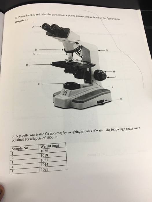
Solved Identify and label the parts of a compound microscope ...
Metaphase - Genome.gov 00:43. Metaphase is a stage during the process of cell division (mitosis or meiosis). Normally, individual chromosomes are spread out in the cell nucleus. During metaphase, the nucleus dissolves and the cell's chromosomes condense and move together, aligning in the center of the dividing cell. At this stage, the chromosomes are ...

National 131-LED-MS Compound Microscope 131-LED-MS B&H Photo
Compound microscope hi-res stock photography and images - Alamy

Parts of a Compound Microscope and Their Functions
Transmitted light microscope B3 Professional series B3-220ASC ...
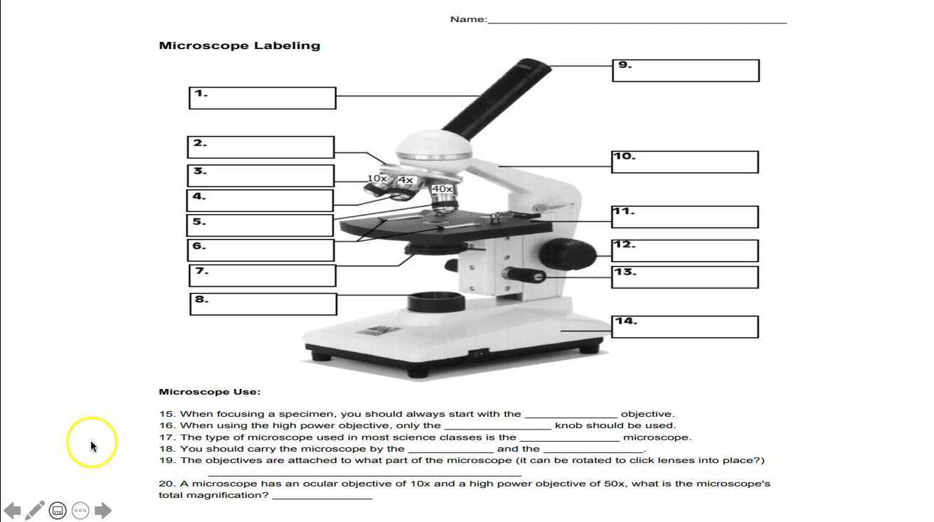
Microscope Labeling

The Compound Microscope Diagram | Quizlet

25 35 Latest Compound Microscope Diagram Ideas - Matahari
Label Parts Of A Compound Microscope Teaching Resources | TpT

Compound Microscope Parts, Functions, and Labeled Diagram ...

Labeling a Compound Microscope Flashcards | Quizlet

E-Katalog 5.0

Simple Microscope - Diagram (Parts labelled), Principle ...

Introductory Hydra Activities

Label Compound Microscope Lesson Plans & Worksheets

What is a Compound Microscope? | Microscope World Blog
Compound Microscope Parts, Functions, and Labeled Diagram ...

Buy National Optical 40X-1000X Compound Microscope Set with ...

Biology - labeling a compound microscope Diagram | Quizlet
What is a compound light microscope? - Dr. Biology Questions ...

2.1 " Compound Microscope" | Download Scientific Diagram

ABOUT MICROSCOPES | Scienceart

Compound Microscope Parts – Labeled Diagram and their ...
Solved Nikon Parts of the compound microscope Write the ...

The Parts of a Compound Microscope and How To Handle Them ...

Compound Microscope Parts – Labeled Diagram and their ...

Microscope Diagram Labeled, Unlabeled and Blank | Parts of a ...

Compound Microscope Parts, Functions, and Labeled Diagram ...

Parts of Stereo Microscope (Dissecting microscope) – labeled ...

microscope binokuler led Oregon di Suntik Cantik vitri | Tokopedia
Post a Comment for "43 label the compound microscope"