41 labeled digestive system diagram
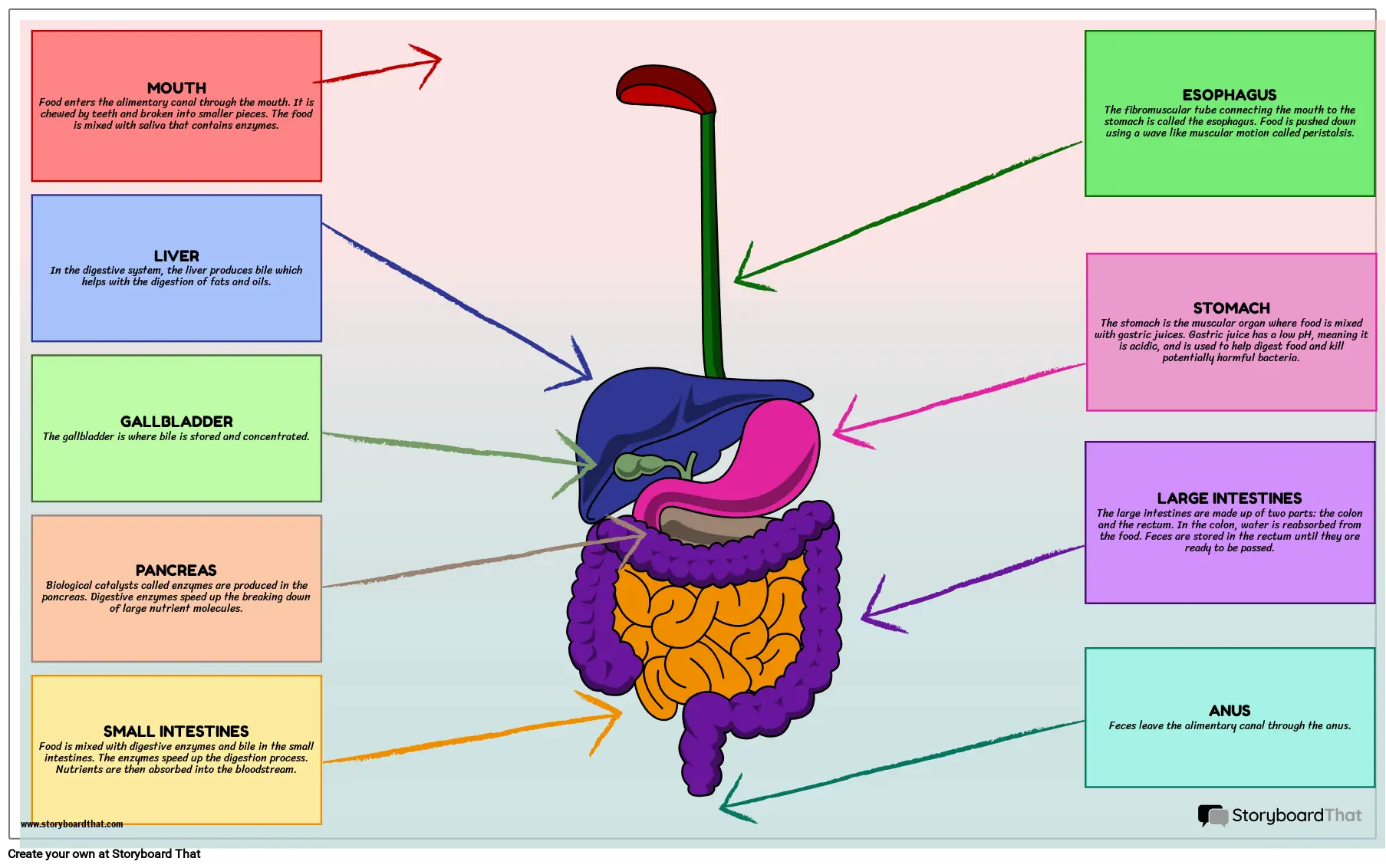
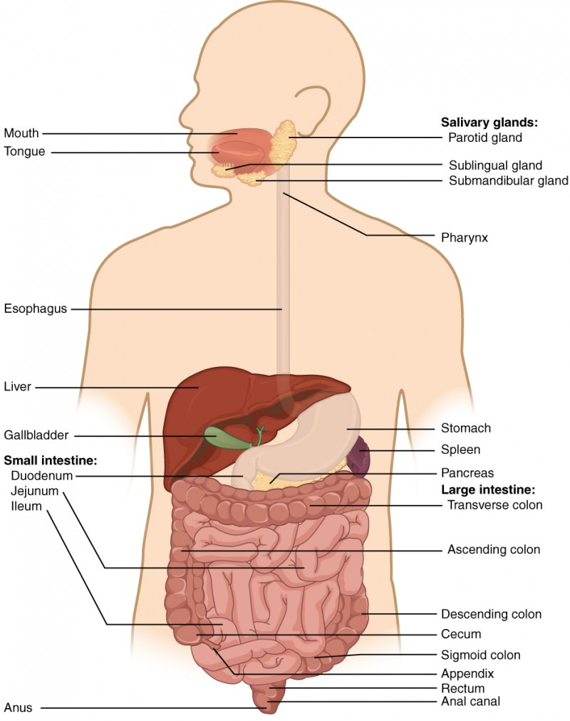
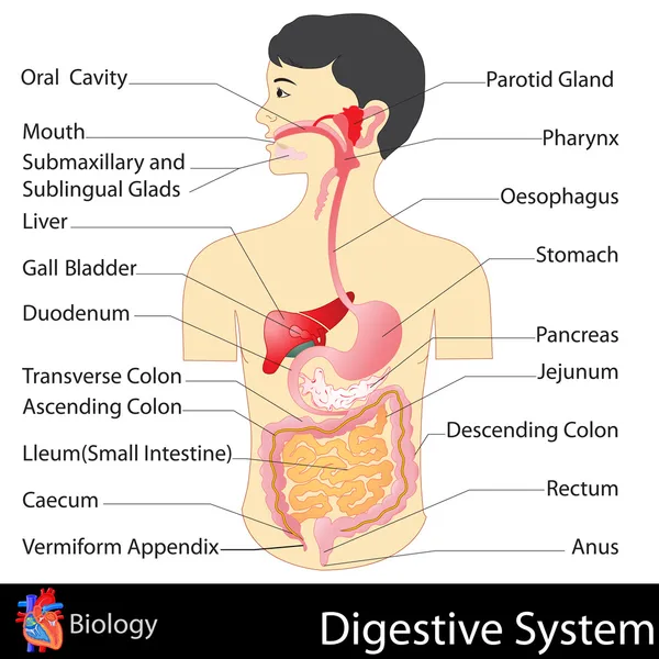
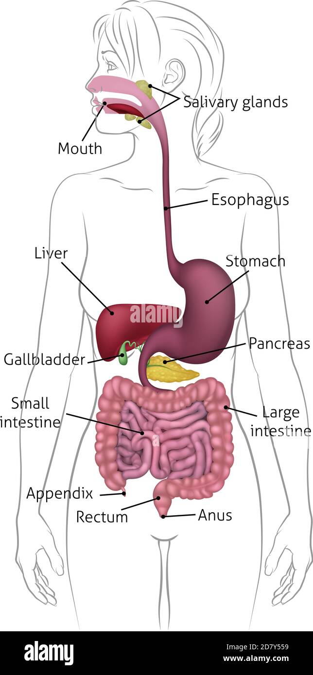
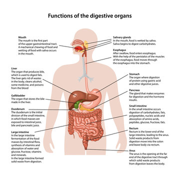
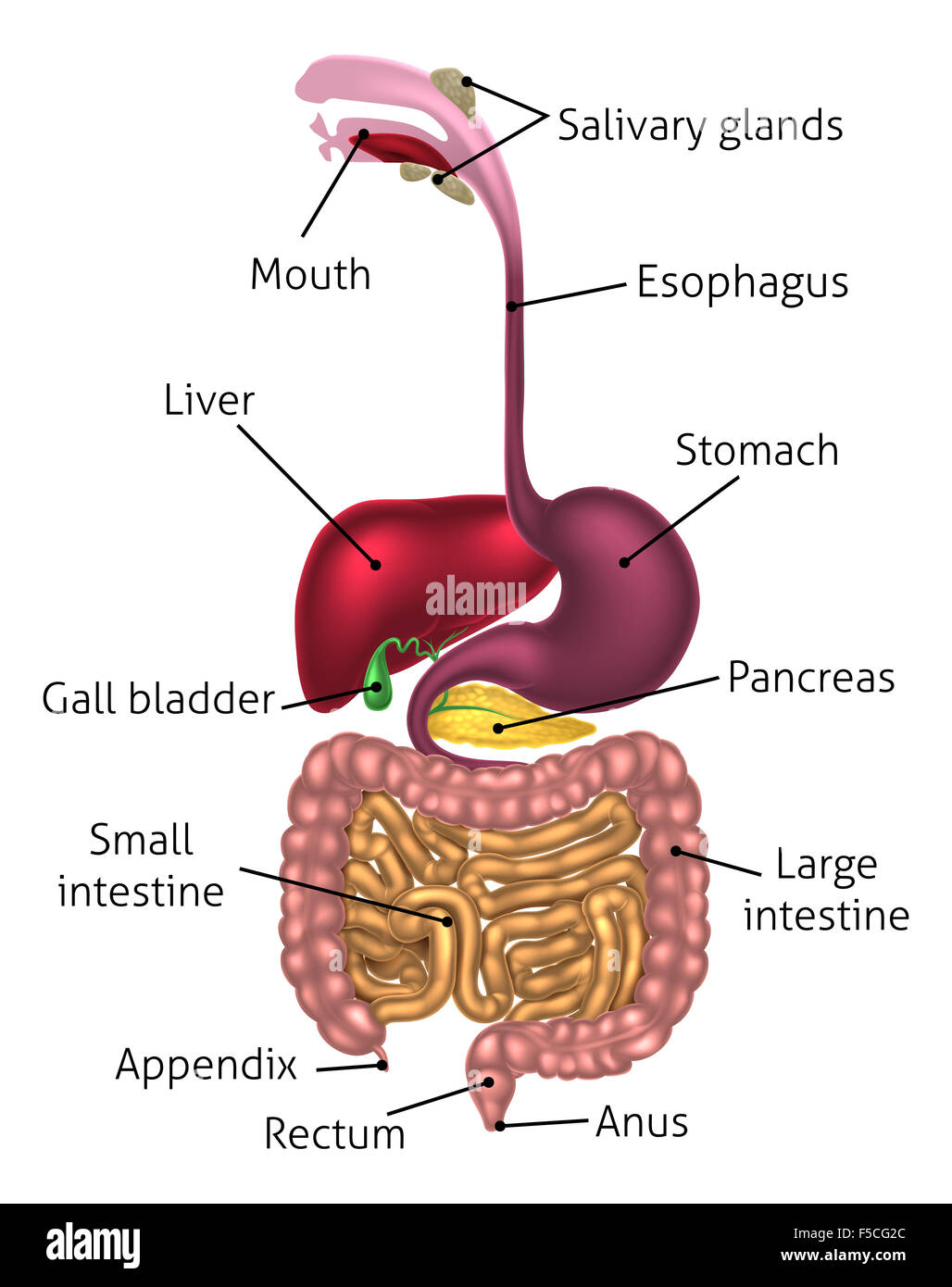
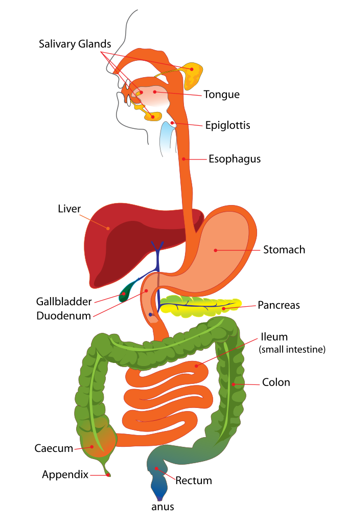
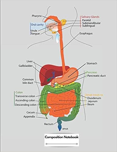
A Labelled Diagram Of Digestive System with Detailed Explanations - Byju's The alimentary canal is divided into five main parts- mouth, esophagus, stomach, small intestine, small intestine and lastly large intestine. The diagram of the human digestive system is useful for both Class 10 and 12. It is one among the few important topics, which are repetitively asked in the board examinations. Also Refer: Alimentary Canal PDF Digestive System Diagram Digestive System Diagram Salivary (food pipe) Liver Large Intestine (ascending colon) Esophagus Tongue Glands Stomach Pancreas Small Intestine (duodenum) Small Intestine (jejunum) Small Intestine (illium) Appendix Anus Large Intestine (transverse colon) Large Intestine (descending colon) Rectum Gallbladder
Digestive System Labeling interactive Quiz - PurposeGames.com This is an online quiz called Digestive System Labeling interactive There is a printable worksheet available for download here so you can take the quiz with pen and paper. From the quiz author Interactive for 5th Graders This quiz has tags. Click on the tags below to find other quizzes on the same subject. digestion Digestive digestive system

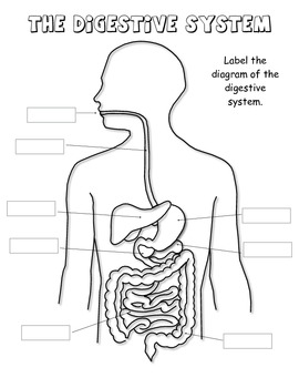
Labeled digestive system diagram
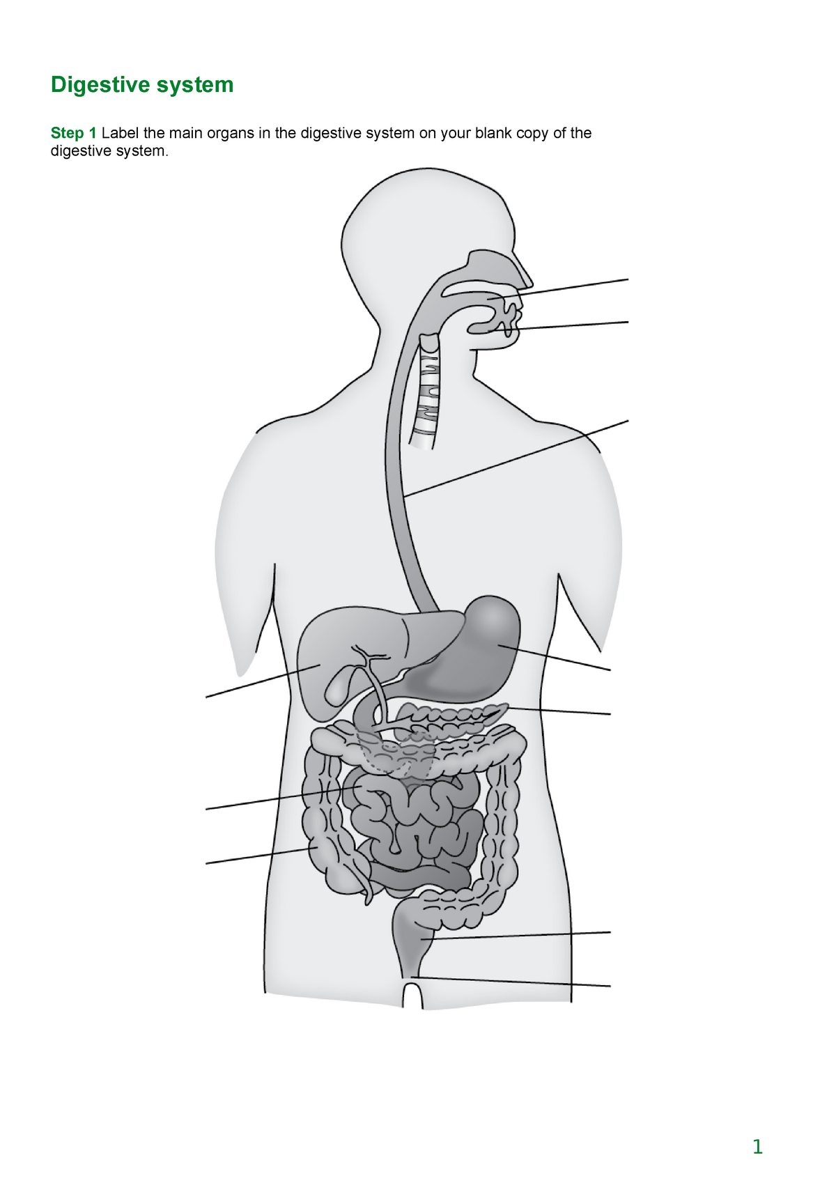
PDF Label the Digestive System - STEP Approach Learning Label the Digestive System Page 2 of 3 Developed by Madeleine Cox Salivary glands Teeth Tongue Epiglottis Oesophagus Trachea Stomach Spleen Diaphragm Liver Gallbladder Small Intestine Pancreas Appendix Large Intestine Rectum . Created Date: The Digestive System Diagram, Organs, Function, and More Accessory Digestive Organs. Pancreas Among other functions, the pancreas is the chief factory for digestive enzymes that are secreted into the duodenum, the first segment of the small intestine ... Digestive System Label Diagram | Quizlet Digestive System Label STUDY Learn Flashcards Write Spell Test PLAY Match Gravity Created by aotineru56 Terms in this set (21) Esophagus - extends from pharynx through diaphragm to stomach - consists of smooth muscles at area close to stomach Small intestine Nutrient absorption - contains enzyme from columnar epithelial cells, pancreas, and liver
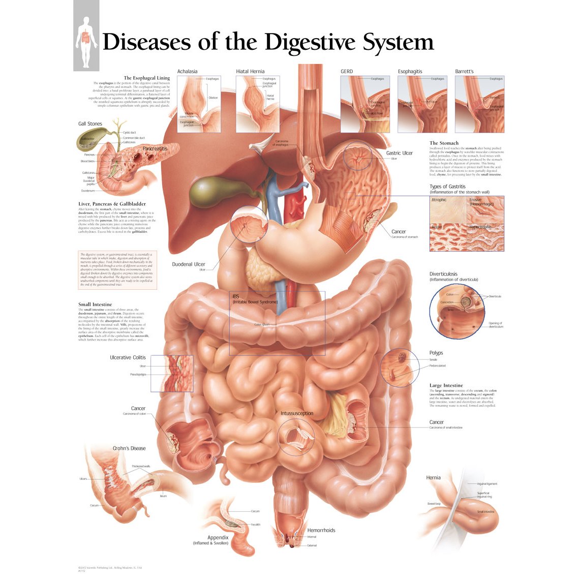
Labeled digestive system diagram. Lymphatic System: Diagram, Function, Anatomy, Diseases The lymphatic system involves many organs, including the tonsils, adenoids, spleen, and thymus. Lymph nodes filter out bacteria and cancer cells and create white blood cells to fight infection. These nodes are found throughout the body (neck, armpits, groin, chest, and abdomen). 1. This article explains the function of the lymphatic system ... Diagram of the Digestive System And an Explanation of its Working Here are some facts about the digestive system: • We produce almost one quart of saliva every day. • Saliva is 98% water and only 2% enzymes. • Length of the digestive tract is about 30 feet. • Food stays in the stomach for almost 2 to 3 hours. • The small intestine is almost 20 feet long. Your Digestive System & How it Works | NIDDK The GI tract is a series of hollow organs joined in a long, twisting tube from the mouth to the anus. The hollow organs that make up the GI tract are the mouth, esophagus, stomach, small intestine, large intestine, and anus. The liver, pancreas, and gallbladder are the solid organs of the digestive system. The small intestine has three parts. Labeled Digestive System Diagram stock illustrations Diagram with superior sigittal sinus, ventricles, arachnoid Villi and spinal cord central canal. labeled digestive system diagram stock illustrations. Tracheostomy cross section diagram, vector illustration Tracheostomy cross section diagram, vector illustration labeled scheme. Intensive care unit equipment medical technology.
the digestive system diagram labeled - ModernHeal.com the digestive system diagram labeled This image is titled the digestive system diagram labeled and is attached to our article about How To Overcome Problems In Your Digestive System. Be sure to visit the guide for more context and information about the digestive system diagram labeled, or read some of our other Health & Anatomy posts! 970 Digestive System Diagram Premium High Res Photos Browse 970 digestive system diagram stock photos and images available, or search for human digestive system or circulatory system to find more great stock photos and pictures. Related searches: human digestive system. circulatory system. Digestive organs: Diagram, stomach, intestines, and more The digestive organs in the abdomen work together to absorb nutrients and move food through the digestion process. They include the stomach, gallbladder, liver, pancreas, intestines, and urinary... Digestive System Diagram - SmartDraw The Human Digestive System Pancreas produces digestive enzymes and bicarbonate solution Stomach stores and breaks down ingested food Gall Bladder stores bile, which aids in digesting lipids Salivary Glands create mucus to aid in digestion Small Intestine (duodenum, jejunum, and ilium) digests carbohydrates, fats, and proteins and absorbs nutrients
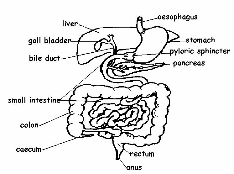
Digestive system labeled Diagram | Quizlet Digestive system labeled Diagram | Quizlet Digestive system labeled STUDY Learn Write Test PLAY Match + − Created by danni_dirato Terms in this set (13) Tongue ... Liver ... Ascending colon ... Cecum ... Appendix ... Rectum ... Trachea ... stomach ... Pancreas ... Transverse colon ... Small intestine ... Descending Colon ... Ileum ... Labeled Digestive System Diagram | 消化器系, 生理学, 人体解剖学 Labeled Digestive System Diagram. Melissa Dunn. 158 followers . Digestive Tract Diagram. Digestive System Parts ... Help students learn and remember the parts of the renal system using this diagram crossword. BONUS ACTIVITY: When they've completed the crossword, get them to cut out the diagram, glue it on a separate page and label the parts of ... Functional Anatomy of the Digestive System: Diagram & Organs The digestive system is made up of the digestive tract. This consists of a long tube of organs that runs from the mouth to the anus and includes the esophagus, stomach, small intestine, and large intestine, together with the liver, gallbladder, and pancreas, which produce important secretions for digestion that drain into the small intestine. Label Digestive System Diagram Printout - EnchantedLearning.com Read the definitions below, then label the digestive system anatomy diagram. anus - the opening at the end of the digestive system from which feces (waste) exits the body. appendix - a small sac located on the cecum. ascending colon - the part of the large intestine that run upwards; it is located after the cecum.
Digestive System Anatomy, Area, and Diagram | Body Maps human body maps digestive system Digestive The human digestive system is the means by which tissues and organs receive nutrients to function. The system breaks down food, extracts nutrients from...
Cat Digestive System Anatomy with a Labeled Diagram Cat digestive system labeled diagram. I know you already got the different labeled diagrams on the cat digestive system organs. Now, it is the final turn for you to identify all the organs from the cat's digestive apparatus with the help of labeled diagrams.
Digestive System - Labelled diagram - Wordwall Digestive System. Share Share by Mstarrfifth. G4 Science Human Body. Show More. Like. Edit Content. Embed. More. Leaderboard. Show more Show less . This leaderboard is currently private. Click Share to make it public. This leaderboard has been disabled by the resource owner. This leaderboard is disabled as your options are different to the ...
Label the digestive system - Labelled diagram - Wordwall mouth/teeth, salivary glands, oesophagus, liver, stomach, small intestine, large intestine, rectum, anus. Label the digestive system Share by Katiemurphy108 KS2 Y4 Science Like Edit Content More Leaderboard Log in required Switch template Interactives
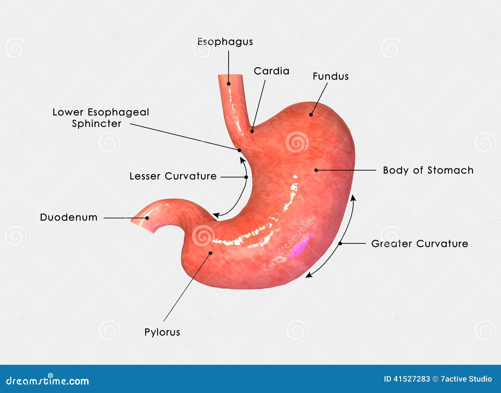
Understanding the Human Stomach Anatomy With Labeled Diagrams Given below is a labeled diagram of the stomach to help you understand stomach anatomy. The stomach is divided into four parts. These include: Cardia Fundus Body Pylorus Cardia refers to the section of the stomach that is located around the cardiac orifice. The lower esophageal sphincter lies at the junction where the esophagus meets the stomach.
Digestive system diagram Stock Photos and Images - Alamy Digestive system diagram Stock Photos and Images. RF PJ459N - Fatty liver disease and hepatic steatosis body part as a medical health care concept of the digestive system anatomy and vital organ for digestion. RM CWTCK7 - Diagram / illustration of the human digestive system drawn on a blackboard in chalk.
Abdomen and digestive system diagrams: normal anatomy | e-Anatomy - IMAIOS This e-Anatomy illustrates the gross anatomy of the digestive system. We focused especially on the diagrams of the abdominal digestive system (oesophagus is described on the modules about the thorax and oral cavity/pharynx on the ENT modules). 84 anatomical diagrams and histological images with over 300 labeled anatomical parts.
Digestive System - The Biology Corner Digestive System. See also Label the Digestive System for practice labeling structurees. For more labeled parts of the human digestive system, view here. This work is licensed under a Creative Commons Attribution-NonCommercial-ShareAlike 4.0 International License.
PDF Label the Digestive System #1 - White Plains Public Schools Label the Digestive System #1 Human Anatomy Read the definitions below, then label the digestive system anatomy diagram. anus - the opening at the end of the digestive system from which feces exit the body. appendix - a small sac located near the start of the large intestine. esophagus - the long tube between the mouth and the stomach. It uses rhythmic muscle
Labeled Diagram Of The Digestive System Stock Photos, Pictures ... Browse 117 labeled diagram of the digestive system stock photos and images available, or start a new search to explore more stock photos and images. Newest results Human Gastrointestinal Digestive System and Labels Medical anatomy illustration of human gastrointestinal digestive system including intestines or gut with labels The digestive system
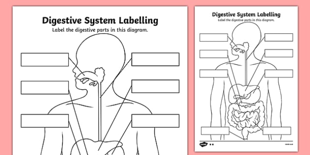
Blank Digestive System Diagram | Labeling Activity - Twinkl This resource contains a diagram of the human body and the digestive system, with spaces to label the names of each internal organ. This resource comes with two differentiated versions - one with a word bank provided and one with blank spaces for your students to complete. An answer key is included for quick grading.
Digestive System Label Diagram | Quizlet Digestive System Label STUDY Learn Flashcards Write Spell Test PLAY Match Gravity Created by aotineru56 Terms in this set (21) Esophagus - extends from pharynx through diaphragm to stomach - consists of smooth muscles at area close to stomach Small intestine Nutrient absorption - contains enzyme from columnar epithelial cells, pancreas, and liver
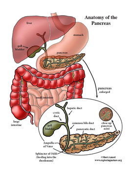
The Digestive System Diagram, Organs, Function, and More Accessory Digestive Organs. Pancreas Among other functions, the pancreas is the chief factory for digestive enzymes that are secreted into the duodenum, the first segment of the small intestine ...
PDF Label the Digestive System - STEP Approach Learning Label the Digestive System Page 2 of 3 Developed by Madeleine Cox Salivary glands Teeth Tongue Epiglottis Oesophagus Trachea Stomach Spleen Diaphragm Liver Gallbladder Small Intestine Pancreas Appendix Large Intestine Rectum . Created Date:















Post a Comment for "41 labeled digestive system diagram"