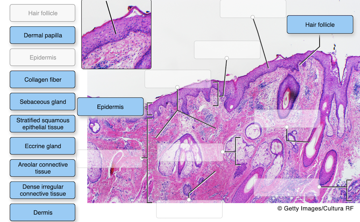
44 label the photomicrograph of the skin and its accessory structures
Solved Label the photomicrograph of the skin and its | Chegg.com Question: Label the photomicrograph of the skin and its accessory structures. Epidermis Sebaceous gland Hair follicle Duct of sebaceous gland Label the photomicrograph of the skin and its accessory structures. Solved Label the photomicrograph of the skin and its | Chegg.com Label the photomicrograph of the skin and its accessory structures. Epidermis Sebaceous gland Hair follicle Duct of sebaceous gland Label the photomicrograph of the skin and its accessory structures. Epidermis Sebaceous gland Hair follicle Duct of sebaceous gland ; Question: Label the photomicrograph of the skin and its accessory structures ...
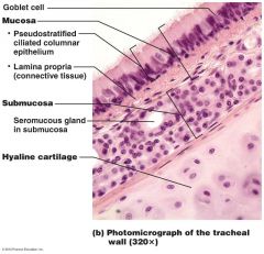
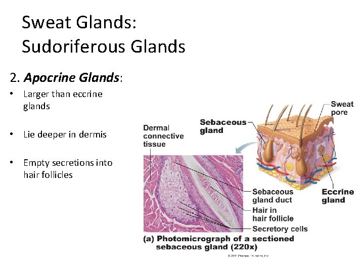
Solved Label the photomicrograph of the skin and its | Chegg.com See the answer Label the photomicrograph of the skin and its accessory structures Epidermis Duct of sebaceous gland Hair follicle Sebaceous gland Show transcribed image text Expert Answer 2. The picture here demonstrates the pseudostratified columnar epithelium.
Label the photomicrograph of the skin and its accessory structures
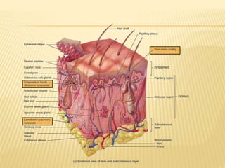
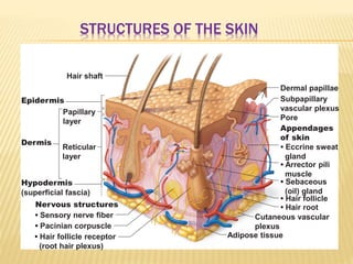
Function And Structure of Skin And Subcutaneous Tissue - Earth's Lab Structure of The Skin And Subcutaneous Tissue. The skin is thickest in areas subjected to wear and tear (abrasion), such as the soles of the feet, where it may be 6 mm in thickness. It is thinnest on the eyelids, eardrums, and external genitalia, where it averages about 0.5 mm in thickness. The skin consists of two major layers: the epidermis ... Integumentary System HW_answers.docx - Course Hero 18.Label the photomicrograph of the skin and its accessory structures. 19.Label the photomicrograph of the sebaceous gland. 20.Label the structures of merocrine sweat glands. 21.Label the structures of the hair follicle. unit 4 lab.docx - LAB Unit 4 EXERCISE 7: The ... - Course Hero Dermis FIGURE 7.4:Diagram of the skin and accessory structures. • apocrine (AP-oh-krin) sweat gland • arrector pili (PIE-lee) muscle • eccrine (EK-rin) sweat gland • hair bulb • hair follicle • hair root • hair shaft • papilla (puh-PILL-uh) of hair • sebaceous (se-BAY-shus) gland 1. Hair shaft 2. Hair root 3. Sebaceous glands 4.
Label the photomicrograph of the skin and its accessory structures. PDF The Integumentary System - Holly H. Nash-Rule, PhD Label the skin structures and areas indicated in the accompanying diagram of thin skin. Then, complete the statements that ... Accessory Organs of the Skin 9. Match the key choices with the appropriate descriptions. Some terms are used more than once. Key: a. arrector pili d. hair follicle g. A&P 1 Exercise_7 Activity 1 & 2 & RYK and UYK.docx - Course Hero Using Your Knowledge A. Identification of Skin from Different Body Locations 7 Observe diagrams of skin from different body locations in Figure 7.7.Based on number of epithelial layers and skin accessory structures, determine if skin is from the axillary area, forearm, or sole of foot. 1. axillary area 2. Sole of foot 3. Forearm B. Application Answer the following questions in the space provided. photomicrograph of thick skin Diagram - Quizlet Start studying photomicrograph of thick skin. Learn vocabulary, terms, and more with flashcards, games, and other study tools. Solved Label the Photomicrograph of the skin and its - Chegg Who are the experts? Experts are tested by Chegg as specialists in their subject area. We review their content and use your feedback to keep the quality high. Transcribed image text: Label the Photomicrograph of the skin and its accessory structures.
Label The Photomicrograph Of The Skin And Its Accessory Structures ... Expert's Answer. The Integumentary System A. Anatomy of the skin. Identify and label the photograph of the skin model: epidermis, dermis, dermal papillae, hypodermis, hair follicle, hair bulb, sebaceous gland, sudoriferous gland, duct of sudoriferous gland, pore of... What are the structures in the integument (skin) which carry responses from ... Solved Label the photomicrograph of the skin and its | Chegg.com Label the photomicrograph of the skin and its accessory structures Epidermis Duct of sebaceous gland Hair follicle Sebaceous gland Show transcribed image text Expert Answer 2. The picture here demonstrates the pseudostratified columnar epithelium. Diagram of human skin structure - Science Learning Hub Diagram of human skin structure. Add to collection. + Create new collection. Tweet. Rights: University of Waikato Published 1 February 2011 Size: 100 KB Referencing Hub media. The epidermis is a tough coating formed from overlapping layers of dead skin cells. Figure 7.1: Photomicrograph of Skin Diagram - Quizlet Start studying Figure 7.1: Photomicrograph of Skin. Learn vocabulary, terms, and more with flashcards, games, and other study tools.
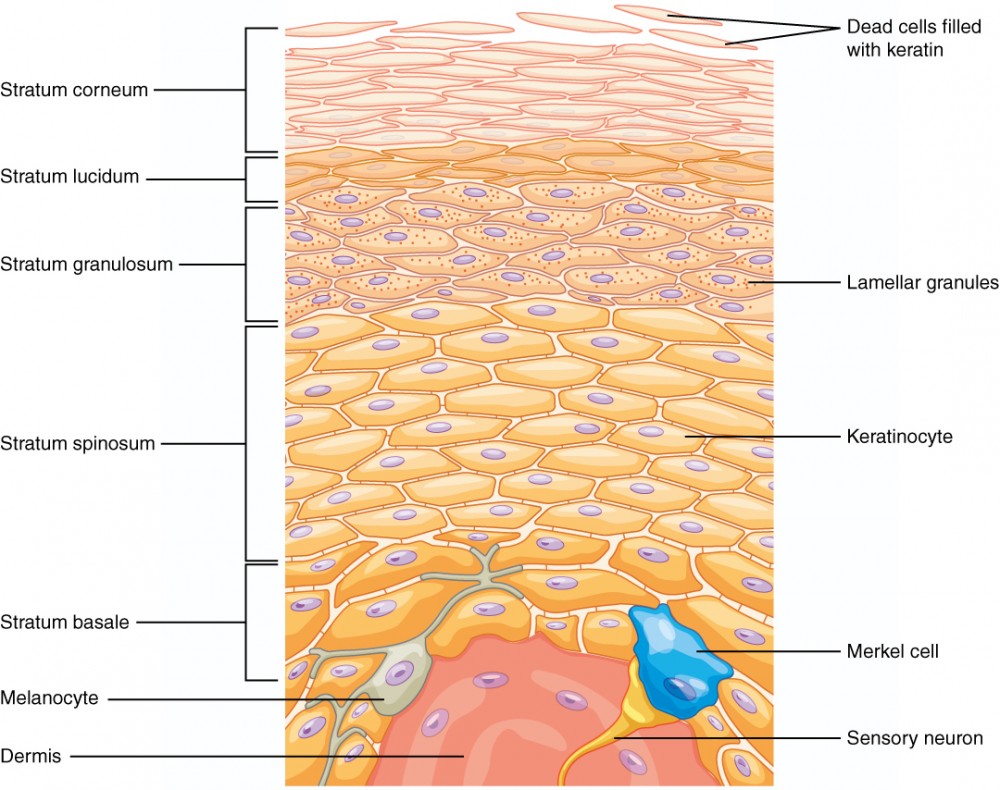
PreLab03a Integument & Prelab03b Integument Histology - Quizlet Categorize the appropriate structures or descriptions in the appropriate layer of skin that is highlighted in blue. Top layer: composed of 5 layers. is avascular. composed of keratinocytes. most superficial layer. Middle layer: composed of 2 layers. contains hair follicles. PDF CHAPTER 5 The Integumentary System - downloads.lww.com tion with its accessory structures. The skin consists of two layers: the epidermis, which includes the outer protective stratum of keratinized epithelial cells, and the dermis, the underlying layer of connective tissue containing blood vessels and nerve endings. The accessory structures include outgrowths of the epidermis (hair and nails). Figure 7.4 Photomicrograph of the skin and accessory structures Start studying Figure 7.4 Photomicrograph of the skin and accessory structures. Learn vocabulary, terms, and more with flashcards, games, and other study tools. RS7 (2).pdf - Basic Structure of the Skin 1. Complete the... View RS7 (2).pdf from GEOL 1402 at Tarrant County College. Basic Structure of the Skin 1. Complete the following statements by writing the appropriate word or phrase on the blank line: 1. The
A & P LAB FINAL Flashcards - Cram.com
Solved Label the photomicrograph of the skin and its 17.01.2020 · Biology. Biology questions and answers. Label the photomicrograph of the skin and its accessory structures. Sebaceous gland Duct of sebaceous gland Epidermis Hair follicle. Question: Label the …
The Integumentary System
Label The Photomicrograph Of The Skin And Its Accessory … 28.03.2022 · Expert's Answer. The Integumentary System A. Anatomy of the skin. Identify and label the photograph of the skin model: epidermis, dermis, dermal papillae, hypodermis, hair follicle, hair bulb, sebaceous gland, sudoriferous gland, duct of sudoriferous gland, pore of...
Integumentary System Overview
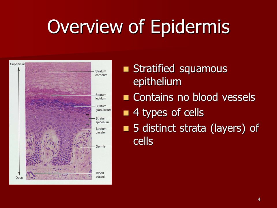
PDF Name the Condition - Dr. Scott Croes' Website the cartoon and the photomicrograph. •Name the Layers of skin and label the dermal papilla and dermis •Name the Layers of skin and label the dermal papilla and dermis. Name the layer of skin shown. Stratum Spinosum. Name the specific layers of skin indicated by the ... Identify the following structures: Epidermis, Hair cortex, Hair medulla ...

PreLab03a Integument & Prelab03b Integument Histology ...
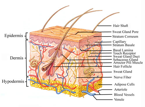
Layers of the Skin - Anatomy and Physiology The skin and its accessory structures make up the integumentary system, which provides the body with overall protection. The skin is made of multiple layers of cells and tissues, which are held to underlying structures by connective tissue (). The deeper layer of skin is well vascularized (has numerous blood vessels).

Ch 4 Skin and Body Membranes Epithelial Membranes
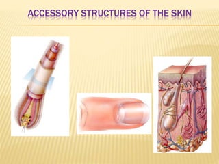
Accessory Structures of the Skin – Anatomy & Physiology Accessory structures of the skin include hair, nails, sweat glands, and sebaceous glands. These structures embryologically originate from the epidermis and can extend down through the dermis into the hypodermis. Hair Hair is a keratinous filament growing out of the epidermis. It is primarily made of dead, keratinized cells.
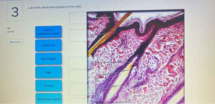
Solved Label the photomicrograph of thin skin 3 10 points ...
Accessory Structures of the Skin - Anatomy & Physiology Accessory structures of the skin include hair, nails, sweat glands, and sebaceous glands. These structures embryologically originate from the epidermis and can extend down through the dermis into the hypodermis. Hair Hair is a keratinous filament growing out of the epidermis. It is primarily made of dead, keratinized cells.
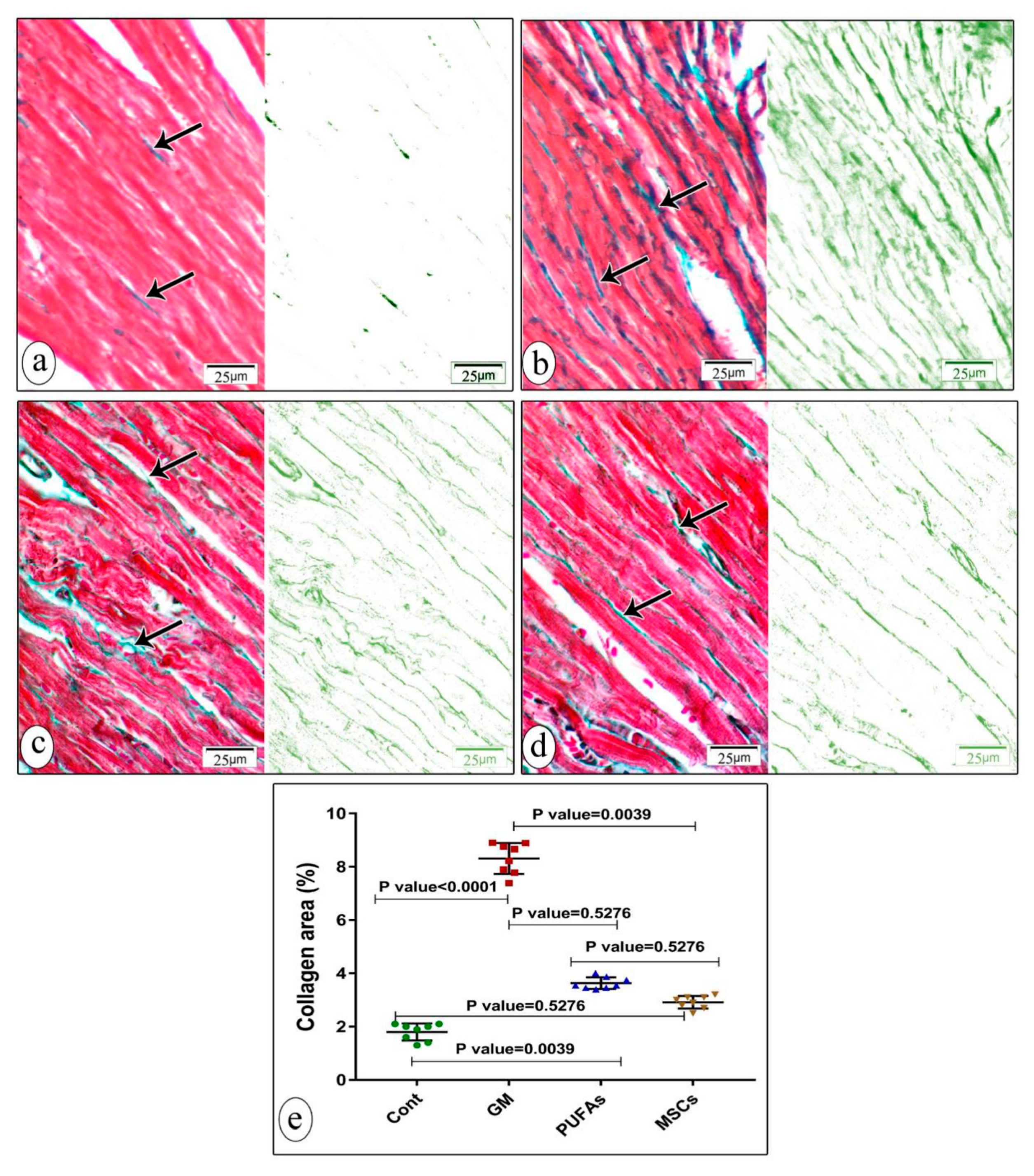
Nutrients | Free Full-Text | Amelioration of Ethanol-Induced ...
Diagram of the skin and accessory structures - Quizlet Start studying Diagram of the skin and accessory structures. Learn vocabulary, terms, and more with flashcards, games, and other study tools.
Flinn Digital Dissection Labs: Pig, 1-Year Access | Flinn ...
Solved Label the Photomicrograph of the skin and its | Chegg.com Transcribed image text: Label the Photomicrograph of the skin and its accessory structures.
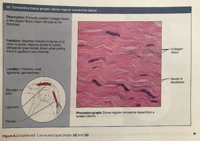
1 Chapter 6 The Integumentary System Skin and its accessory ...
Skin 2: accessory structures of the skin and their functions All are important in the skin's key functions, including protection, thermoregulation and its sensory roles. This article, the second in a two-part series, looks at the structure and function of the main accessory structures of the skin. Citation: Lawton S (2020) Skin 2: accessory structures of the skin and their functions.

Photomicrograph of the cleared head of a juvenile channel ...
Figure 7.4 Photomicrograph of the skin and accessory structures Start studying Figure 7.4 Photomicrograph of the skin and accessory structures. Learn vocabulary, terms, and more with flashcards, games, and other study tools.

Exercise 8: photomicrograph of spongy bone Flashcards | Quizlet
Laboratory Exercise 4 Fillable .pdf - NEW ERA UNIVERSITY... Identify the accessory structures of the skin on models or microscope slides. 4 Describe the function of the epidermal accessory structures. 5 Compare eccrine sweat gland density on the forehead, forearm, palm, and anterior leg. ... Label the photomicrograph in Figure 4.4. 3.
Chapter 5
Label the photomicrograph in Figure 7.4. Examine a slide of hairy skin ... Expert's Answer Solution.pdf Next Previous Label the layers of the skin on the diagram and the photograph. Be able to identify the layers on a microscope slide. Look at the skin slide under a microscope. a) Epidermis 1) Stratum corneum ii) Stratum lucidum 111) Stratum granulosum iv) Stratum... Recent Questions in Basics of Statistics Q:
Layers of the Skin | Anatomy and Physiology I
Solved Label the photomicrograph of the skin and its | Chegg.com Question: Label the photomicrograph of the skin and its accessory structures. Sebaceous gland Duct of sebaceous gland Epidermis Hair follicle This problem has been solved! See the answer Show transcribed image text Expert Answer 100% (19 ratings)
A Comparative Account of the Morphological and Anatomical ...
Answered: 1. In the photomicrograph below of… | bartleby Science Anatomy and Physiology Q&A Library 1. In the photomicrograph below of cartilage tissue, find and label the indicated structures. Extra cellular r Lacuna Chondrocyte Dyte Elastic protein fibers Extracellular matrix In the photomicrograph below of compact bone tissue, find and label the indicated structu p Osteon Lamella Lacuna o Osteocyte Canaliculi Central canal
Lab Exam 2 Exercise 6&7 Flashcards | Chegg.com
Figure 7.1: Photomicrograph of Skin Diagram | Quizlet Start studying Figure 7.1: Photomicrograph of Skin. Learn vocabulary, terms, and more with flashcards, games, and other study tools.

Chapter 6 Study Set Flashcards | Quizlet
Solved Label the photomicrograph of thick skin | Chegg.com Anatomy and Physiology questions and answers; Label the photomicrograph of thick skin ; Question: Label the photomicrograph of thick skin . This problem has been solved! See the answer See the answer See the answer done loading. Show transcribed image text Expert Answer. Who are the experts?
Integumentary System – Building a Medical Terminology Foundation
Anatomy, Skin (Integument), Epidermis - StatPearls - NCBI Bookshelf Skin is the largest organ in the body and covers the body's entire external surface. It is made up of three layers, the epidermis, dermis, and the hypodermis, all three of which vary significantly in their anatomy and function. The skin's structure is made up of an intricate network which serves as the body's initial barrier against pathogens, UV light, and chemicals, and mechanical injury ...

Damaged Skin | SpringerLink
pg 87 fig 7.4 diagram of skin and accessory structures - Quizlet Start studying pg 87 fig 7.4 diagram of skin and accessory structures. Learn vocabulary, terms, and more with flashcards, games, and other study tools.

unit 4 lab.docx - LAB Unit 4 EXERCISE 7: The Integumentary ...
PreLab03a Integument & Prelab03b Integument Histology - Quizlet Label the structures of the skin and subcutaneous tissues. Left side form top: ... stratum spinosum stratum basale. Categorize the appropriate structures or descriptions in the appropriate layer of skin that is highlighted in blue. Top layer: ... Label the photomicrograph of the skin and its accessory structures. epidermis hair follicle duct of ...

Integumentary system Module 3: Accessory Structures of the ...
unit 4 lab.docx - LAB Unit 4 EXERCISE 7: The ... - Course Hero Dermis FIGURE 7.4:Diagram of the skin and accessory structures. • apocrine (AP-oh-krin) sweat gland • arrector pili (PIE-lee) muscle • eccrine (EK-rin) sweat gland • hair bulb • hair follicle • hair root • hair shaft • papilla (puh-PILL-uh) of hair • sebaceous (se-BAY-shus) gland 1. Hair shaft 2. Hair root 3. Sebaceous glands 4.

PDF) Comparison of mitochondrial transplantation by using a ...
Integumentary System HW_answers.docx - Course Hero 18.Label the photomicrograph of the skin and its accessory structures. 19.Label the photomicrograph of the sebaceous gland. 20.Label the structures of merocrine sweat glands. 21.Label the structures of the hair follicle.

anatomy lab, exam 3, lab 9, Spinal Nerves, Integument, and ...
Function And Structure of Skin And Subcutaneous Tissue - Earth's Lab Structure of The Skin And Subcutaneous Tissue. The skin is thickest in areas subjected to wear and tear (abrasion), such as the soles of the feet, where it may be 6 mm in thickness. It is thinnest on the eyelids, eardrums, and external genitalia, where it averages about 0.5 mm in thickness. The skin consists of two major layers: the epidermis ...
Integumentary System Flashcards - Easy Notecards
Chapter 5

Solved label the diagram of the skin and accessory | Chegg.com
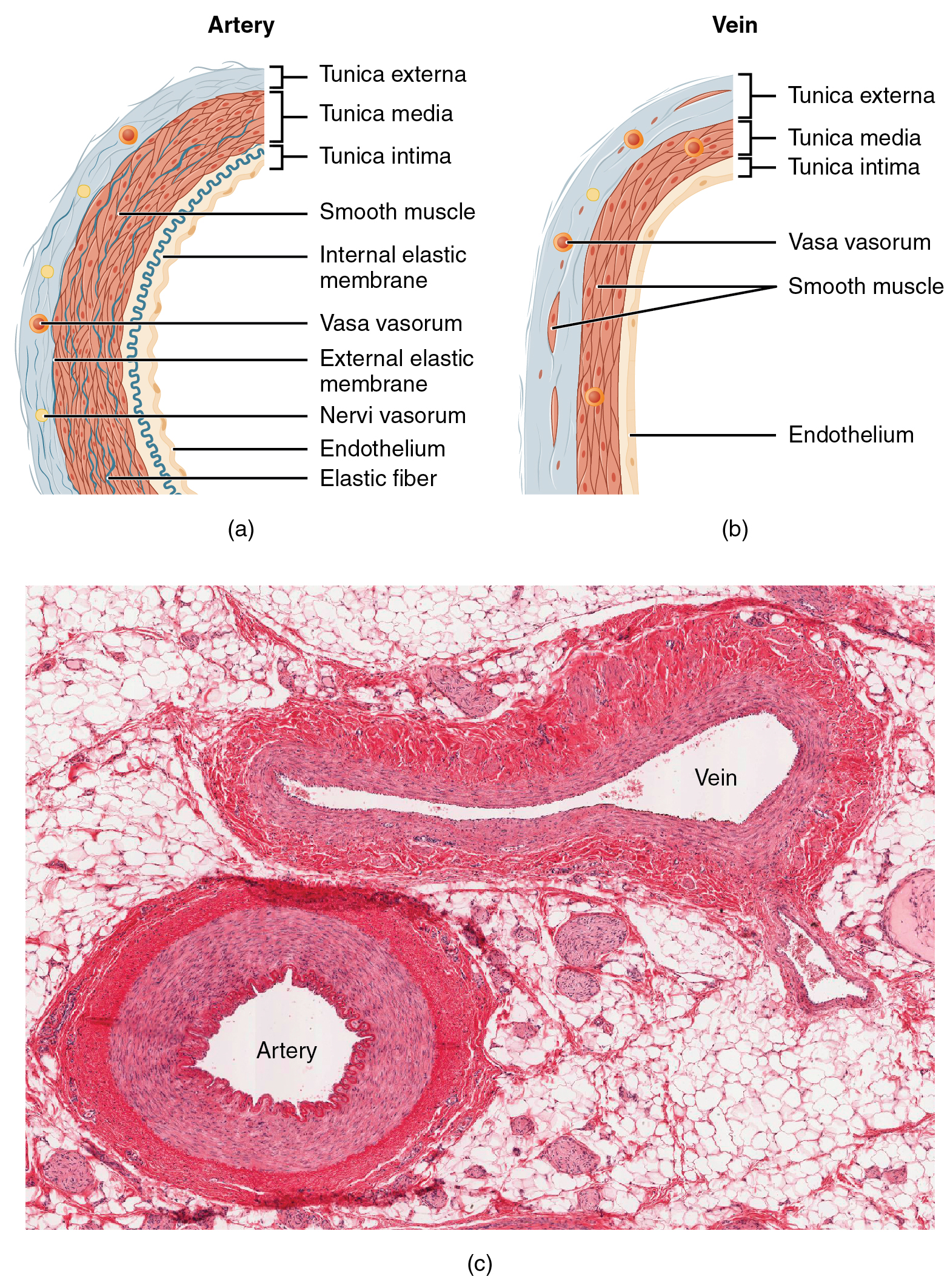
Structure and Function of Blood Vessels – Anatomy and Physiology

Photomicrograph of rat epididymis stained with HE (× 200 ...

unit 4 lab.docx - LAB Unit 4 EXERCISE 7: The Integumentary ...

A & P lab test 4 Flashcards | Quizlet
Solved Label the structures of the skin in this micrograph ...
Vital Pulp Extirpation
Pharmaceutics | Free Full-Text | Therapeutic Potential of ...

Chapter 15 - Accessory Structures of the Skin - BIO 140 ...

JaypeeDigital | eBook Reader

Photomicrographs of Nissl-stained transverse sections showing ...

Exploring Molecular Mechanisms of Aloe barbadmsis Miller on ...
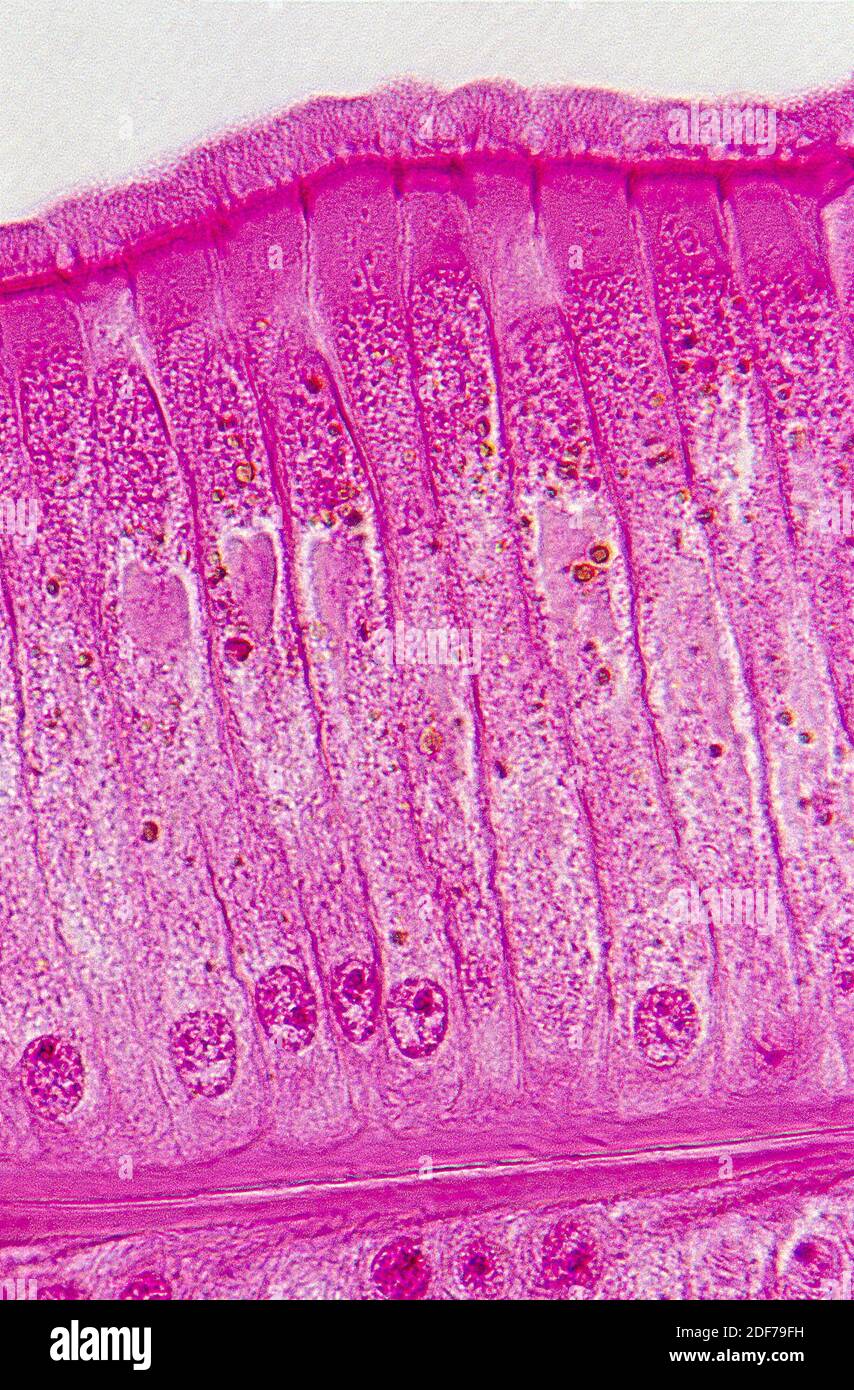
Simple ciliated columnar High Resolution Stock Photography ...
The Integumentary System

Histo Exam 3 FINAL EXAM Flashcards | Quizlet

Photomicrograph of rat epididymis stained with HE (× 200 ...

Layers of the Skin | Anatomy and Physiology I
Integumentary System 2 Regions 1 Epidermis 2 Dermis

Anatomy of the Lymphatic and Immune Systems | Anatomy and ...
Chapter 5
Post a Comment for "44 label the photomicrograph of the skin and its accessory structures"